Restorative Only vs. Gingivectomy + Restorative: A Visual Treatment Planning Case Study
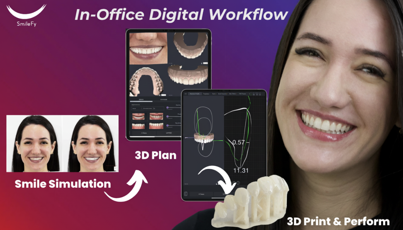
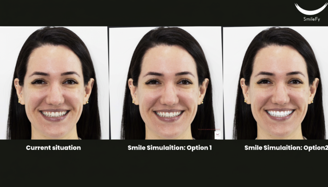
Do you agree that Visual Treatment Planning plays a tremendous role in Patient Decision-Making? In this case study, we explore the decision-making process of a patient with a high lip line and short laterals and premolars. Initially, the patient came in wanting to increase the length of the upper laterals only — however, when we created different smile simulations to show the patient a visual of different esthetic outcomes, her perspective changed dramatically.
1. Patient Profile
The patient presented with a chief complaint of short upper lateral incisors and a desire to improve overall smile aesthetics. Clinical examination and smile analysis identified the following key features relevant to treatment planning:
High lip line — the patient displayed excessive gingival tissue on full smile — a finding that is relevant to both the restorative and periodontal components of the treatment options.
Short maxillary lateral incisors and premolars — the clinical crown length of the lateral incisors and premolars was reduced relative to ideal proportions, with excess gingival tissue covering part of the anatomical crown.
Initial treatment request — the patient originally requested lengthening of the upper laterals only — a restorative-only starting point that the planning process subsequently expanded to include a full options comparison.
The patient's decision was significantly influenced by a chairside try-in of both planned outcomes — illustrating how digital planning and physical mockups directly support informed patient consent and treatment acceptance.
2. Imaging and Planning Data
The following data were collected to support 3D digital smile design and to enable precise planning of the gingivectomy option:
Facial photographs — full-face frontal smile photographs taken in Natural Head Position (NHP) for facial analysis and smile frame alignment.
Intraoral scan (IO scan) — upper arch intraoral scan providing the 3D tooth and gingival surface geometry used for smile design overlay and silicone key fabrication.
CT scan (CBCT) — cone-beam computed tomography providing bone crest data for the posterior aesthetic zone. The CBCT was used with SmileFy's real-time cross-section function to plan the gingivectomy with precision, verify biologic width safety, and determine whether osteoplasty (bone reduction) would be required alongside soft tissue excision.
In cases where the planned gingival margin is close to the alveolar bone crest (less than 3 mm), a CT scan is required to determine whether soft tissue removal alone is sufficient or whether osseous contouring (osteoplasty) is also needed. Planning without bone data risks violating biologic width, leading to chronic inflammation, bone resorption, and gingival recession post-operatively.
3. Options Compared
Two complete treatment approaches were designed, modelled, and physically tried in the patient's mouth before a final decision was made.
Composite resin restorations in the aesthetic zone to add length and improve proportions without periodontal surgery.
Gingivectomy and osteoplasty to expose greater clinical crown length, followed by aesthetic composite veneers.
Adequate attached gingiva above planned margins; biologic width not at risk; patient prefers non-surgical approach; low-to-moderate lip line.
High lip line with visible gingival excess; short clinical crowns due to altered passive eruption; CBCT confirms adequate bone-to-margin distance (>3 mm).
Non-surgical. Topical/local anaesthesia for composite placement. No healing period.
Surgical — local anaesthesia; gingivectomy incision ± osseous recontouring. Healing period required.
None — restorations placed at the same appointment.
Soft tissue only: 4–8 weeks. With osteoplasty: 3–6 months for full stabilisation.
Crown lengthening limited to composite thickness. Excess gingival display remains. Risk of bulky restorations.
Surgical margin repositioning of 1.5–3+ mm apically. Gingival display reduced proportionally. Ideal crown proportions achieved.
Composite: 5–10 years. Margin stability inherent — no periodontal healing required.
Periodontal outcome stable long-term if biologic width respected. Composite: 5–10 years. More durable aesthetically.
Over-contouring risks plaque retention and gingival inflammation. Biologic width encroachment if margins too subgingival.
Post-op discomfort, swelling, temporary sensitivity. Rare: insufficient attached gingiva, margin creep.
Standard composite maintenance: polishing, staining monitoring, periodic replacement.
Periodontal maintenance 3–6 months post-surgery. Standard composite maintenance thereafter.
4. Patient Selection and Contraindications
How a high lip line influences the decision: A high lip line is the single most important patient characteristic determining whether the restorative-only approach will produce an acceptable esthetic outcome. When the patient displays more than 2–3 mm of gingival tissue on full smile, composite additions to the incisal edge will lengthen the crown without reducing the gingival display — the smile will continue to look gummy.
The gingivectomy + restorative option directly addresses the high lip line by repositioning the gingival margin apically, reducing gingival display on full smile, and simultaneously correcting the crown proportion.
How short laterals and premolars influence the choice: Short lateral incisors and premolars in the context of a high lip line typically indicate altered passive eruption (APE) — the gingival margin has not fully receded to its final anatomical position, leaving excess tissue covering part of the anatomical crown.
In this scenario, the restorative-only approach would add composite to teeth that are already at the correct incisal length for the face. Gingivectomy exposes the existing anatomical crown, after which the composites can be placed to refine shape and proportion rather than to compensate for tissue excess.
When restorative-only risks biologic width violation: If composite margins are placed subgingivally to achieve the desired crown length — extending 1–2 mm beneath the free gingival margin — they encroach on the junctional epithelium and connective tissue attachment zone (biologic width: approximately 2 mm total). Violation of this zone triggers an inflammatory response, bone resorption, and gingival recession.
5. Step-by-Step Workflow
Step 1 — Data Capture: Capture facial photograph in NHP. Take full-arch intraoral scan. Acquire CBCT of the upper jaw centred on the aesthetic zone (voxel size ≤ 0.2 mm recommended). Time: approximately 30–45 minutes.
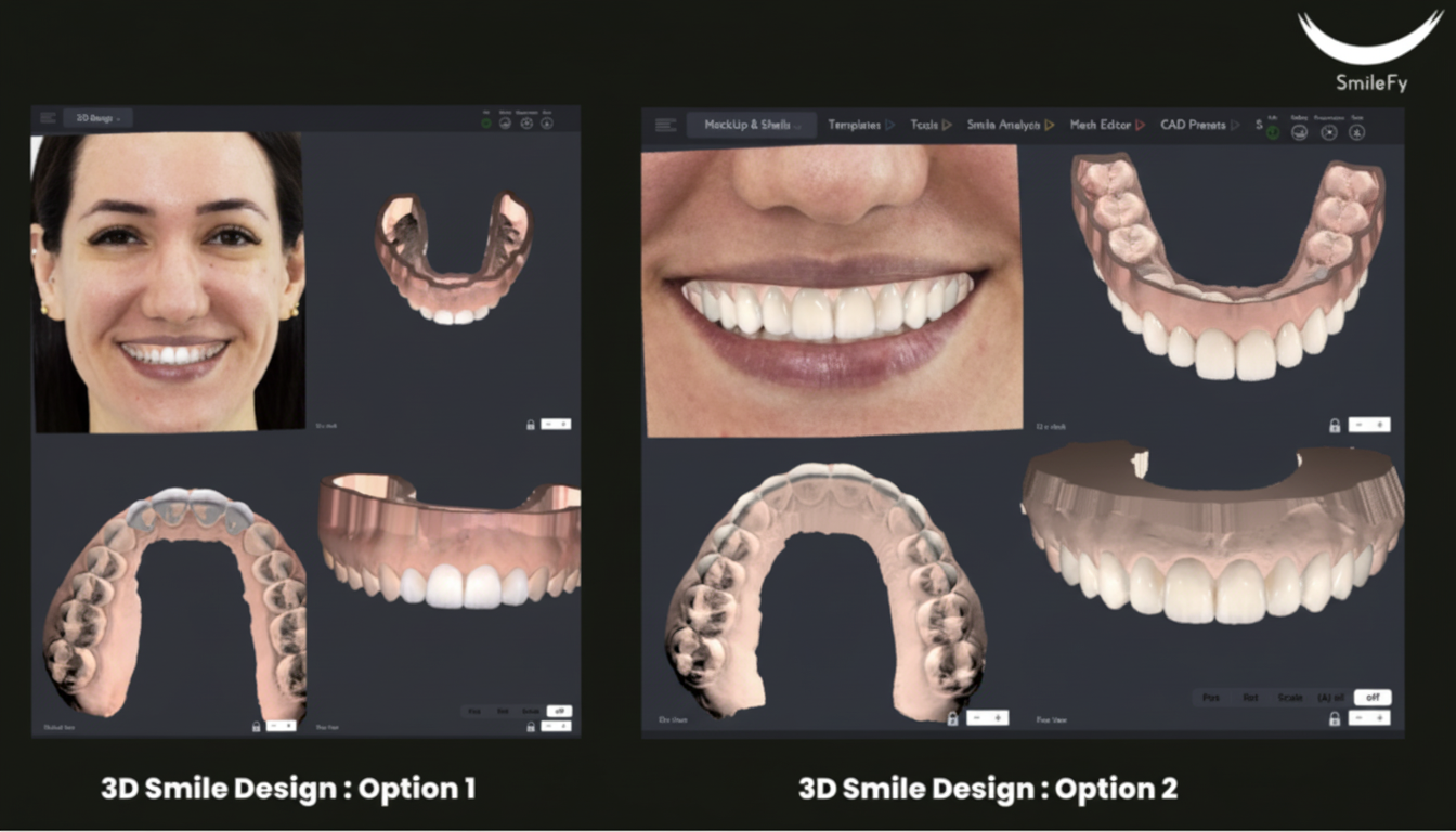
Step 2 — 3D Smile Design (Two Versions): Design Version A (Restorative only): overlay composite additions on the existing gingival architecture using the tooth library. Design Version B (Gingivectomy + Restorative): use the CBCT cross-section to plan the gingivectomy margin level, verify biologic width safety (planned margin ≥ 3 mm from bone crest), then design composite veneers on the repositioned tissue architecture. Time: approximately 60–90 minutes for both designs.
Step 3 — 3D Print Physical Models: Export the STL files for both designs from SmileFy. Print a physical model for each design. Each model represents the planned tooth form and gingival margin position for its respective treatment option. Print time: typically 1–3 hours per model.
Step 4 — Fabricate Silicone Keys: Press or vacuum-form a silicone index over each printed model to capture the planned crown form. These silicone keys are used chairside to inject composite resin into the correct position and shape for each intraoral mockup. Time: approximately 15–30 minutes per key.
Step 5 — Chairside Try-In (Both Options): At a dedicated try-in appointment, seat the silicone key for Version A and inject mockup material. Allow to set. Remove the key and evaluate the restorative-only outcome in the patient's mouth. Photograph for documentation. Repeat with the silicone key for Version B to show the planned post-surgical aesthetic result. Time: approximately 60 minutes for both try-ins.
Step 6 — Patient Decision: After evaluating both try-ins in her mouth and reviewing photographs, the patient selected Option B: gingivectomy and osteoplasty in the posterior aesthetic zone, followed by aesthetic composite veneers.
Step 7 — Surgical Appointment: Seat the silicone guide to mark the planned new gingival margins. Perform gingivectomy to the marked levels. Where the CBCT cross-section indicated insufficient bone-to-margin clearance (< 3 mm), perform osteoplasty to restore the biologic width before tissue excision. Appointment length: typically 60–90 minutes.
Step 8 — Healing Period: Soft tissue gingivectomy: 4–8 weeks for margin stability before restorations. With osteoplasty: 3–6 months for complete hard and soft tissue stabilisation.
Step 9 — Definitive Restorations: Place the composite veneers using the same silicone key approved during the try-in. The key ensures the definitive restoration matches the shape and position the patient approved.
Step 10 — Follow-Up: Review appointment at 6–8 weeks post-restoration, then at 6 months. Evaluate gingival health at restoration margins, composite integrity, patient satisfaction, and occlusal stability.
6. Risks and Aftercare
Surgical risks (gingivectomy + osteoplasty):
Post-operative discomfort and swelling — expected in the first 24–72 hours; managed with standard analgesics and ice.
Temporary tooth sensitivity — exposed root surfaces or newly uncovered enamel may exhibit thermal sensitivity during healing.
Margin creep / recession — if biologic width was underestimated or violated, the gingival margin may recede further apically after healing.
Insufficient attached gingiva — aggressive excision in patients with a narrow band of attached gingiva may leave < 2 mm of attached tissue.
Restorative risks (both options):
Marginal staining and secondary caries — composite margins require monitoring and periodic polishing.
Chipping or fracture — patients with parafunctional habits (bruxism) have elevated risk; a night guard is indicated.
Biologic width violation (restorative-only) — placing subgingival margins restoratively without periodontal crown lengthening risks triggering chronic tissue inflammation and bone resorption.
Aftercare instructions:
Post-surgical — soft diet for 48–72 hours; gentle rinsing with 0.12% chlorhexidine or saline; no flossing in the surgical site until instructed.
Post-restorative — avoid staining beverages for 48 hours post-placement; standard oral hygiene maintained; night guard fabricated if bruxism is present or suspected.
Periodontal maintenance — visits at 3 months post-surgery, then at 6-month intervals for the first year.
7. Outcomes
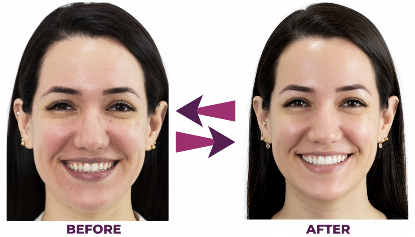
Following the gingivectomy and osteoplasty, the patient's gingival margins were repositioned to the levels planned digitally in SmileFy. The composite veneers were subsequently placed using the silicone key derived from the 3D printed model, ensuring that the final restorations matched the design the patient had approved during the try-in appointment.
Patient decision confidence — the chairside try-in of both treatment options was the primary factor enabling the patient to make an informed decision between the two approaches.
Precision from digital planning — the CBCT real-time cross-section in SmileFy allowed precise pre-surgical identification of the extent of osteoplasty required, reducing intraoperative decision-making.
Aesthetic outcome — the final composite veneers were placed on a correctly proportioned tissue architecture — crown length and gingival margin positions were established surgically, with the restorations refining form and shade.
8. Key Takeaways
Visual treatment planning changes treatment acceptance — presenting digital smile simulations and physical intraoral mockups enables patients to select between treatment options based on direct experience rather than clinician description alone.
High lip line + short laterals/premolars = altered passive eruption until proven otherwise — CBCT is essential to distinguish APE from true crown shortness.
Restorative-only is a compromise when gingival excess is the primary cause — composite additions to the incisal edge do not reduce gingival display; only margin repositioning does.
CBCT cross-section planning prevents biologic width violations — pre-surgical bone-to-margin distance measurement distinguishes soft tissue-only gingivectomy cases from those requiring osteoplasty.
Staged workflow protects the restorative outcome — placing definitive composite margins before gingival margin stability is confirmed wastes chair time and materials.
The silicone key bridges digital design and chairside execution — the same key used for the try-in guides the definitive restoration.
9. Frequently Asked Questions
Q: How long does healing take after a gingivectomy before restorations can be placed?
Soft tissue gingivectomy only: allow 4–8 weeks for gingival margin stability; with osteoplasty: 3–6 months for complete hard and soft tissue stabilisation before placing definitive restorations.
Q: How uncomfortable is the gingivectomy procedure and recovery?
The procedure is performed under local anaesthesia and is not painful during surgery; post-operative discomfort (soreness and mild swelling) is expected for 2–5 days and is typically manageable with standard over-the-counter analgesics and soft diet.
Q: How long will the composite veneers last?
With good home care and regular monitoring, direct composite veneers typically last 5–10 years before refinishing, repair, or replacement is indicated; longevity is influenced by occlusal loading, oral hygiene, and dietary habits.
Q: What maintenance does this treatment require long-term?
Periodontal maintenance visits at 3–6 month intervals for the first year post-surgery; standard composite maintenance (polishing at hygiene visits, monitoring for marginal staining and integrity); a night guard if bruxism is present; and routine dental hygiene appointments thereafter.
Q: Can a restorative-only approach ever achieve the same outcome?
In cases where the lip line is low-to-moderate and crown shortness is genuinely due to incisal deficiency rather than gingival excess, restorative-only can produce excellent outcomes; in high lip line cases with altered passive eruption, the gingivectomy + restorative approach is more appropriate.
Q: Why was a CBCT scan necessary for this case?
The CBCT provided bone crest data that allowed pre-surgical determination of whether osteoplasty was required alongside the soft tissue excision to respect biologic width — information that cannot be reliably obtained from probing alone in all cases.
How this post will look when shared
smilefy.com
Restorative Only vs. Gingivectomy + Restorative: A Visual Treatment Planning Case Study
How digital smile design and chairside try-ins helped a patient with a high lip line choose between restorati…