You're Scanning Every Patient. You're Designing From Photos. Something Doesn't Add Up.
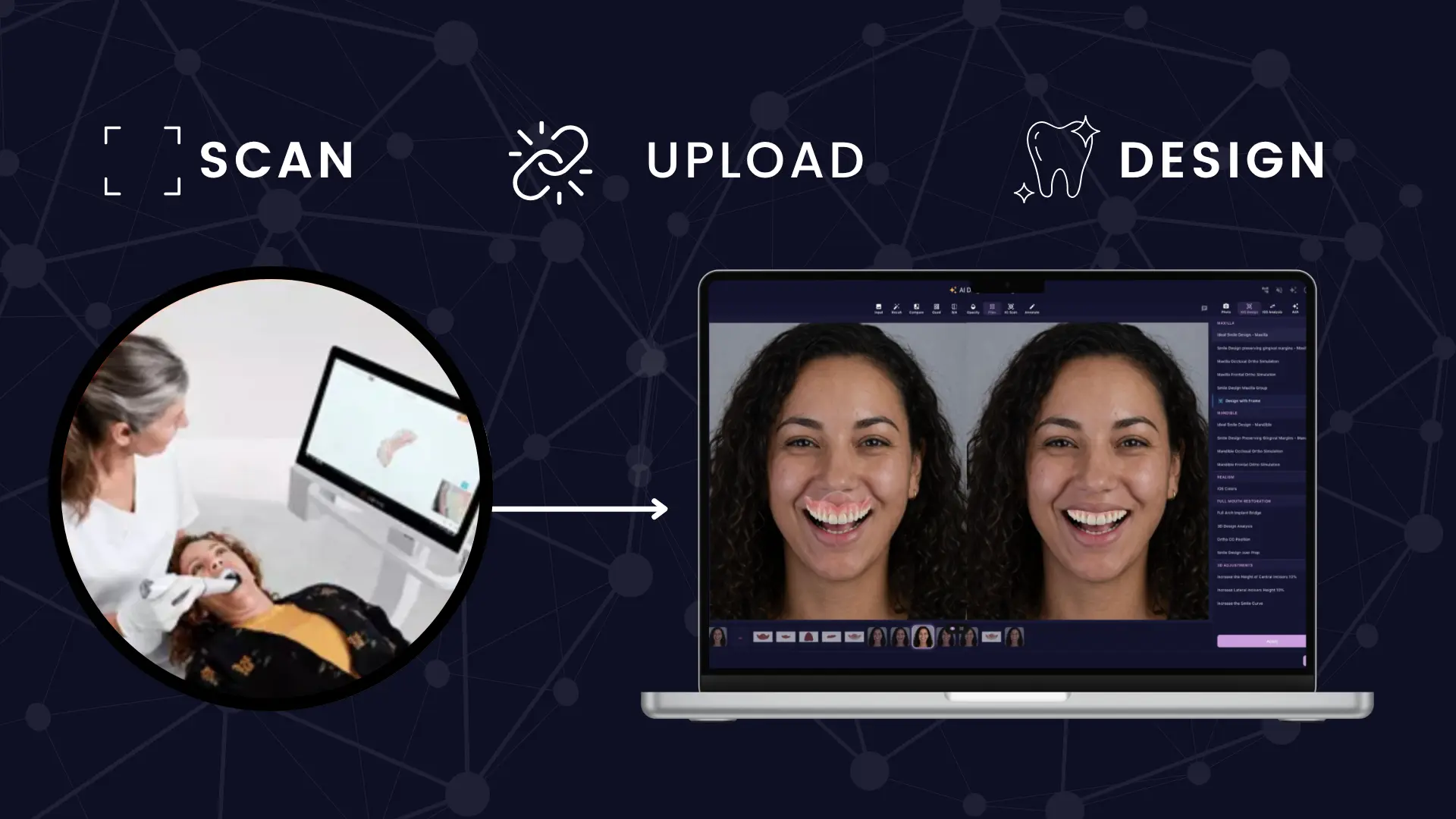
Intraoral scanner adoption just crossed 60% in the U.S. and Northern Europe. Most of those practices made the switch for the same reasons — cleaner impressions, no more trays, patients who arrive expecting it. The scanner earned its place in the workflow quickly.
What it hasn't earned yet, in most practices, is its place in esthetic treatment design.
The same device that captures a complete, dimensionally accurate 3D model of your patient's dentition every time you use it is sitting one software connection away from generating veneer designs, diagnostic wax-ups, and full-arch treatment plans — without a lab planning cycle. In most practices, that connection has never been made.
That's not a gap in clinical knowledge. It's a gap in workflow. And it's narrower than most practices realise.
A Photo Shows You What a Smile Looks Like. A Scan Tells You What It Is.
Photo-based esthetic design works. A well-composed facial photograph, correctly lit and taken with adequate resolution, can produce a simulation credible enough to move a patient from curiosity to commitment. For the consultation stage, a photo is sufficient.
For treatment design, the story is different.
A photograph is a projection — light reflected from surfaces, captured in two dimensions. It records appearance. It does not record tooth geometry, preparation margins, occlusal topography, or the three-dimensional spatial relationships that determine whether a veneer design will seat, function, and integrate with the patient's existing dentition over time.
When a clinician designs from a photo, they are working against a visual target. The prep is planned against the dentist's experience and judgment rather than against a verified 3D record of what is actually there. In uncomplicated anterior cases with experienced hands, this produces good outcomes routinely. In cases with occlusal complexity, multiple units, or minimal preparation, the distance between the visual target and clinical reality is exactly where unpredictability enters.
A scan removes that distance before the first bur touches enamel.
The Research That Most Practices Haven't Seen
There is a documented hesitancy among clinicians about using intraoral scanners for veneer cases specifically. The concern is clinically grounded — veneer preparations present smaller, more delicate margins than full-coverage restorations, often positioned at or sub-gingivally, where scan accuracy could theoretically be compromised.
The literature has now addressed this directly enough to settle the question.
A 2025 narrative review in the *Journal of Clinical Medicine*, covering the published evidence from 2010 to 2025, concluded that intraoral scanning is a reliable and clinically acceptable technique for veneer impressions — with workflow efficiency and chairside time reduction as documented benefits alongside accuracy. The same review cited a large retrospective study reporting cumulative survival of 99.83% for lithium disilicate CAD/CAM veneers over several years. A separate systematic review confirmed that marginal adaptation of CAD/CAM veneers is clinically equivalent to conventionally fabricated restorations.
Technique matters. A 2024 study in the *Journal of Esthetic and Restorative Dentistry* established that scan pattern affects accuracy for complete-arch laminate veneer cases — and that modifying that pattern according to preparation design measurably improves results. That is a learnable protocol adjustment, not a fundamental limitation of the technology.
What the evidence consistently shows is that the hesitancy most practices carry toward scan-based veneer design is not a response to clinical data. It is a response to unfamiliarity. Those are different problems with different solutions.
The Same Case, Two Workflows
The difference between a photo-based and scan-based esthetic workflow is easier to understand when it is mapped against a specific case. A six-unit anterior veneer case is a useful reference point because it is common enough to be recognisable and complex enough for the differences to matter.
Photo-based: Facial photographs taken → 2D simulation generated and approved by patient → case forwarded to lab with photos and written clinical instructions → lab interprets the design intent → diagnostic wax-up returned two to three weeks later → try-in and adjustments → final fabrication proceeds.
The wax-up in this sequence is the first moment the three-dimensional reality of the proposed design becomes visible. Adjustments here are routine — not because anything went wrong, but because the design was never grounded in actual 3D tooth geometry. The lab has been interpreting a communication document, not executing against a clinical dataset.
Scan-based: Facial photographs taken alongside IO scan → AI generates 3D treatment design from scan data calibrated to facial esthetic parameters → design reviewed chairside with the patient → print-ready STL produced → diagnostic mockup printed in-office or delivered to lab as precise dimensional instructions → try-in confirms what was already designed to the patient's actual anatomy.
The try-in in the second sequence is not a discovery moment. It is a confirmation. The 3D design was verified against the patient's actual scan before any physical output was produced. Surprises at this stage are less frequent because the design was accurate from the start rather than approximate.
Both paths can produce excellent clinical outcomes. The number of steps, the margin for interpretive error between intent and execution, and the elapsed time between consultation and confirmed design are not equivalent.
When the Scan Makes the Biggest Difference
Experienced clinicians produce good photo-based esthetic work every day. There is no argument here that scan-based design is required for every case. The argument is that it changes the clinical equation in specific situations — and that those situations represent a significant portion of the esthetic caseload in most practices.
Multi-unit anterior cases. Six to ten veneers redesigning the anterior zone simultaneously involve three-dimensional tooth relationships that a photo cannot capture. Incisal edge positions, gingival contours, and axial inclinations across the full arch are spatial data. Designing against a facial photograph addresses them visually. Designing against a scan addresses them dimensionally. For complete-arch esthetic work, that distinction shows up at the try-in stage.
Cases with occlusal involvement. Any case that changes vertical dimension, modifies an edge-to-edge relationship, or requires careful management of the posterior occlusion while altering anterior geometry cannot be fully planned from a frontal photograph. A 2D simulation captures nothing about what is happening behind the canines. A scan captures everything.
Minimal and no-prep veneers. The smaller the preparation, the higher the precision requirement for the design that precedes it. When there is no reduction to compensate for inaccuracies in the design, the design has to be right before the case begins. Scan-based design provides the dimensional accuracy that makes minimal-prep cases predictable. Photo-based design makes them aspirational.
In-office printing workflows. A printable STL file has to be generated from actual tooth geometry. There is no route from a 2D photo simulation to a print-ready file without rebuilding the design in a separate environment. A scan-based design produces the STL directly. If in-office printing is part of how you deliver esthetic treatment — or if it's something you're considering — the scan is the only starting point that works.
Why the Connection Hasn't Been Made
The scanner penetration curve and the CAD adoption curve in U.S. dentistry are not moving together. Sixty percent of practices now have an intraoral scanner. Only 38% use CAD/CAM design systems. Among the practices that haven't adopted CAD/CAM, 62% cite complexity — not cost — as the reason.
That gap has been stable for years. It is not closing because the scanner got better or the evidence got clearer. It is stable because the software that connects scan data to esthetic treatment design has historically required training, staffing infrastructure, and clinical bandwidth that most practices haven't had.
The scanner sitting in your operatory is ready for this workflow. The missing piece has always been software that can receive scan data and produce a treatment design without asking the clinician to become a CAD operator first.
What Changes When You Connect the Two
SmileFy accepts intraoral scan data from 3Shape TRIOS, Medit, Dexis, AllieStar, and any other scanner via standard import — and generates complete 3D treatment designs without CAD expertise at any stage. The clinician uploads the scan, selects treatment parameters, and SmileFy's AI produces a design calibrated to both the scan geometry and the patient's facial esthetics from the consultation photographs. No CAD environment. No design specialist. No separate planning software.
The output is a print-ready STL file. It can go directly to an in-office printer, or it can be shared with a dental lab as precise dimensional instructions rather than a visual communication document. Either way, what the lab or printer receives is a design grounded in the patient's actual anatomy — not an interpretation of a photograph.
For patient communication, the same platform generates the 2D simulation from photos in seconds. The consultation visual and the clinical treatment design exist in the same workflow, produced from the same patient data, without any rebuilding between them.
That is the connection the scanner was always capable of supporting. The workflow to use it is available without new hardware, a CAD training requirement, or a change to the lab relationship.
The Question That Follows
Most practices that invested in an intraoral scanner made a good decision. The impression quality improved. The patient experience improved. The scanner earns its place at every appointment it's used for.
The question worth sitting with is whether the 3D dataset it captures — accurate, complete, available within minutes of a patient sitting down — is being used for the highest-value clinical decisions that dataset could support.
A photo-based esthetic workflow is not wrong. It is a ceiling. The scan data your practice is already collecting is the foundation for something more dimensionally accurate, more predictable at the delivery stage, and more connected from consultation through to production than a 2D simulation can be.
The ceiling is not the scanner's limitation. It is the software connection that was never made.
---
SmileFy offers a 7-day free trial with full access — no credit card required. The most direct test is to run a current esthetic case through the scan-based workflow and compare what comes out to your existing design process.
Sources
*Accuracy of Digital Impressions for Veneer Restorations: A Narrative Review and Case Illustration*, Journal of Clinical Medicine, 2025
*Effect of preparation design and scan pattern on the accuracy of intraoral scans for complete arch laminate veneers*, Journal of Esthetic and Restorative Dentistry, 2024
*Accuracy of Intraoral Scanner Systems for Fabricating Inlay, Onlay, and Veneer Restorations: A Systematic Review and Meta-Analysis*, Journal of Esthetic and Restorative Dentistry, 2025
*3Shape Digital Dentistry Trends Report*, 2026
Jarvis Analytics: CAD/CAM adoption and complexity barrier data
How this post will look when shared
smilefy.com
You're Scanning Every Patient. You're Designing From Photos. Something Doesn't Add Up.
Intraoral scanner adoption just crossed 60%, but most practices still design esthetic cases from 2D photos. H…