Pre-Surgical Digital Aesthetic Planning for Immediate Loading Screw-Retained Dental Implants
Dr. Adrian Granella
SmileFy Member — G.Clinic, Mendoza, Argentina
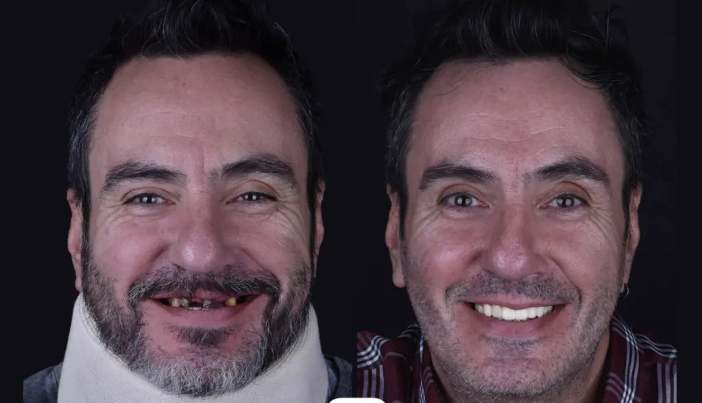
Patient Summary
Introduction
The aesthetic planning of screw-retained dental prostheses is essential in the pre-surgical stage. It helps the clinician to obtain a customized facially guided esthetic and functional planning of the future prosthesis, facilitates the implant planning position, to subsequently install natural and facially balanced dental prosthesis. However, due to its challenging steps, many dentists lapse in this process, often done analogically.
To overcome the limitations of analog workflows, a new dental technology allows clinicians to work better on their cases, helping them to collaborate efficiently with their dental technicians and ultimately deliver higher predictability on their final outcomes to their patients using SmileFy Software.
This case report aims to show the importance of implementing Pre-Surgical Digital Aesthetic Planning of immediate loading screw-retained prostheses to assist dental surgeons in developing faster and more satisfactory final results with their dental technicians in a digital environment.
The Case
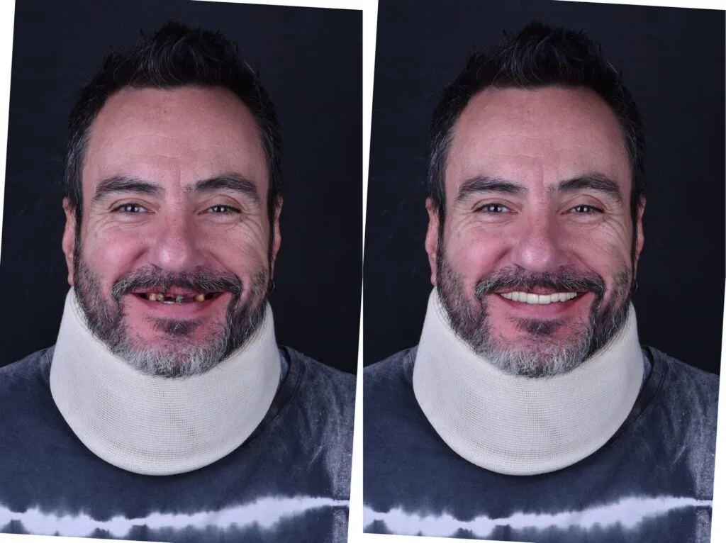
A mid-aged male patient came to Dr. Adrian Granella at the G.Clinic — Mendoza, Argentina, for a dental consultation with severe oral problems.
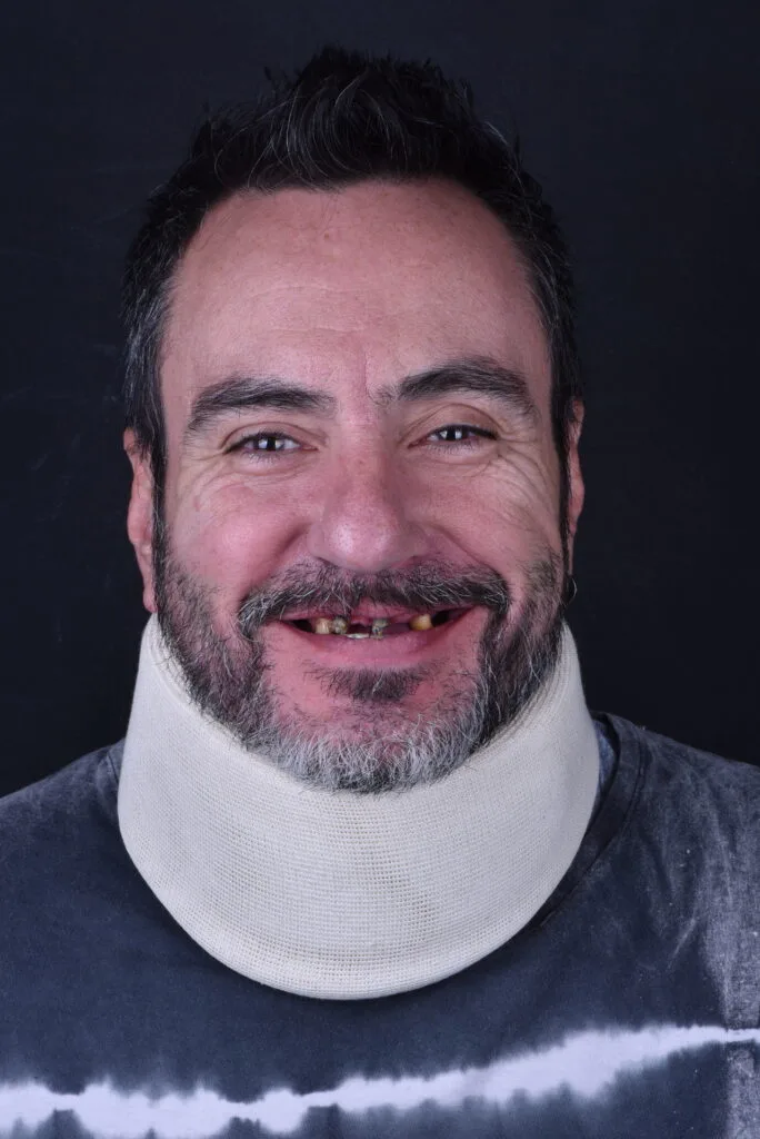
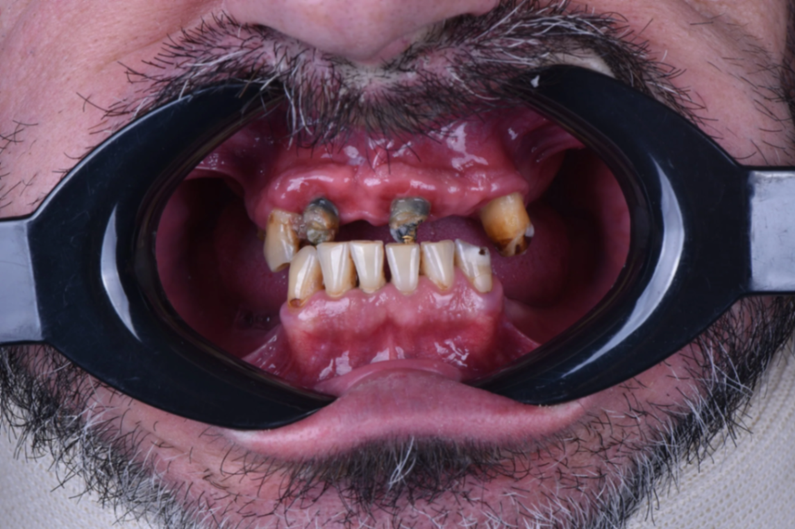
During the clinical examination, the absence of various teeth and compromised carious crowns and roots on both the upper and lower arch, loss of vertical dimension, bone loss, and absence of occlusal plane were observed. This condition left the patient in social and emotional distress.
Although a full-mouth rehabilitation using a full upper screw-retained bridge over implants and single crowns over implants on the lower posterior arch was planned, the patient first chose to start with the upper arch.
Uploading the Digital Files
To digitalize the patient for pre-surgical upper digital bridge design, upper and lower intraoral scans and registration of the new VDO, facial photographs, and CT scans were taken.
3D Dental Bridge Design and Pre-Surgical Esthetic Planning Evaluation
The software allows dentists to superimpose the patient's photograph and digital casts to digitally pre-design the implant bridge while evaluating teeth contour, smile curvature, and proportions, facilitating communication between the interdisciplinary team and patient towards a more incredible rehabilitative therapy and predictable outcome.
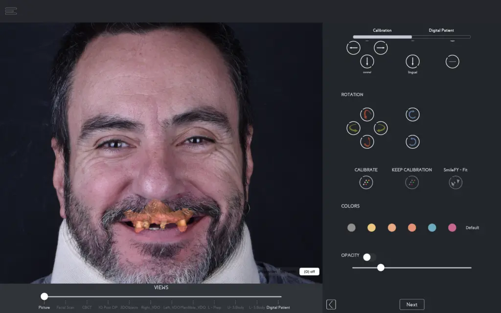
With the files calibrated, the automated 3D smile frame allows the dentist to preset the proportions and position of the entire upper arch and occlusal angulation virtually, which can be fully customized according to the patient's palate form, interocclusal space, and planning.
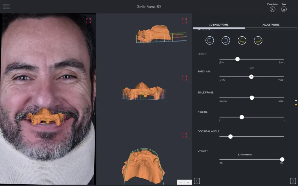
Once the 3D frame is preset, the dental professional can select how many units will be added to the dental bridge, and the 3D teeth templates will automatically appear according to the smile frame setup.
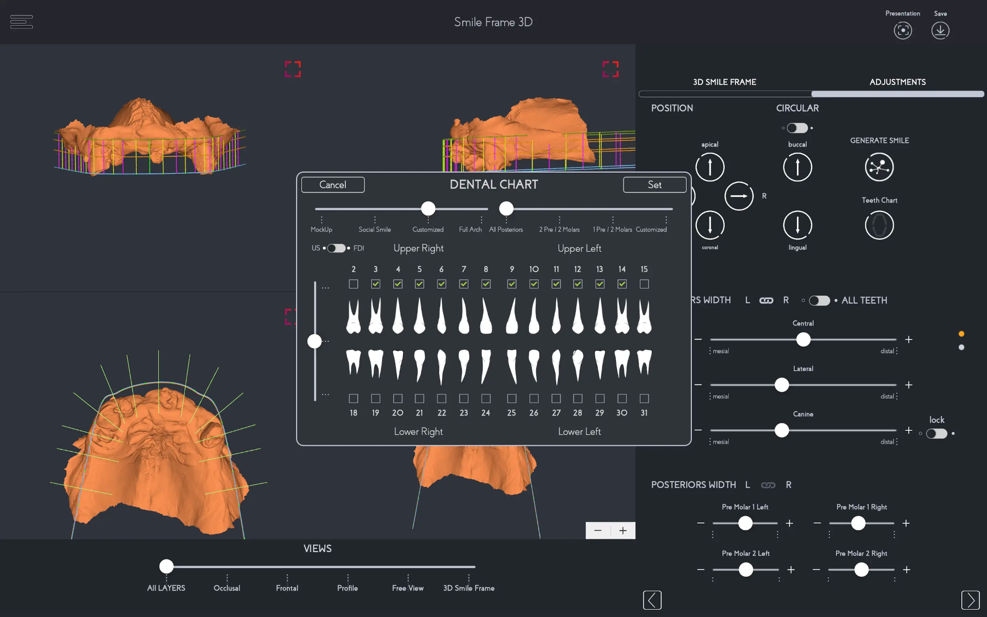
3D Bridge Design
With the 3D teeth automatically generated into the patient's virtual mouth, the dental professional can select from a range of natural 3D teeth templates that can later be exported and shared with the dental technician. The shape selection was based on the patient's desires which facilitates the patient's comfort in participating in the planning stage.
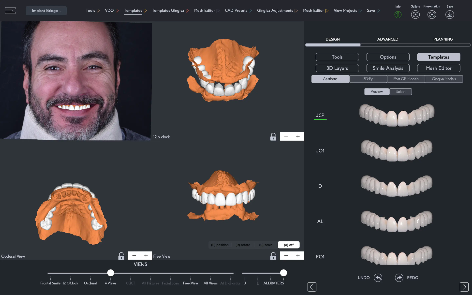
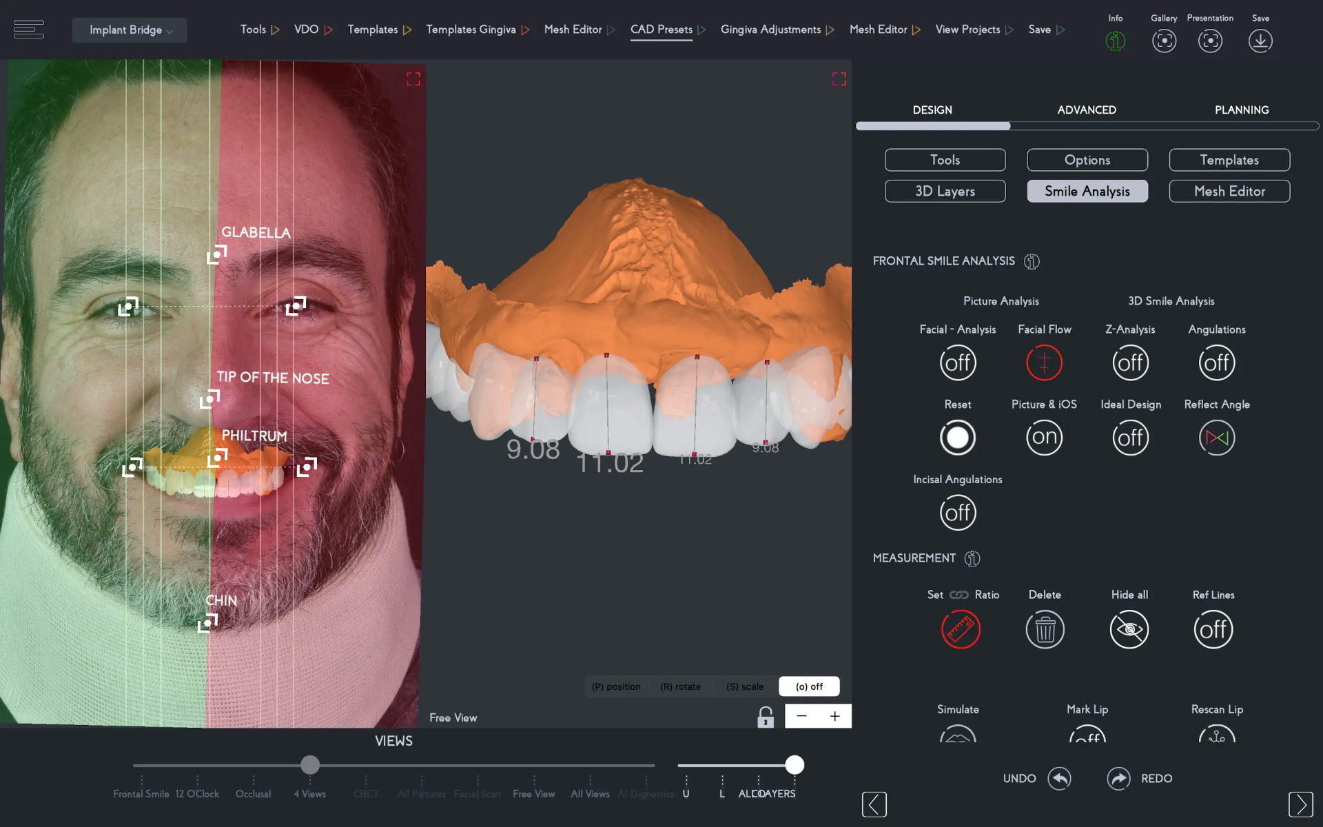
An important step in the pre-surgical esthetic planning process is the virtual evaluation of the bridge design in relation to the patient's face. In the Smile Analysis section of SmileFy, dental professionals can:
Facial midline evaluation — Make corrections to the midline of the prosthesis if needed
Cant assessment — Evaluate the cant of the prosthesis in accordance with the patient's interpupillary and commissural lines, automatically detected through A.I. technology
Smile curvature — Evaluate the anterior smile curvature line of the dental bridge to refrain from creating an inverted smile
Teeth proportions — Measure the teeth' height and width to respect the principles of esthetics in smile design
With the teeth template in place, the artificial gingiva is added with one click and customized in the mesh editor feature.
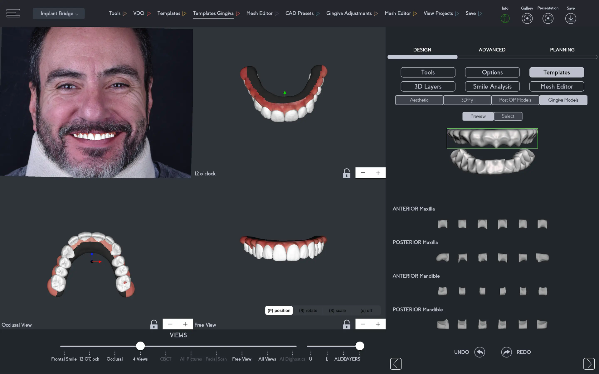
In this stage, the dental professional can customize the dental bridge esthetics to boost the communication between the dentist and lab technician regarding gingival contouring, palatal base, and more, as well as to pre-plan the position of implants according to the patient's arch form and treatment planning when sending the implant bridge design to an implant planning software.
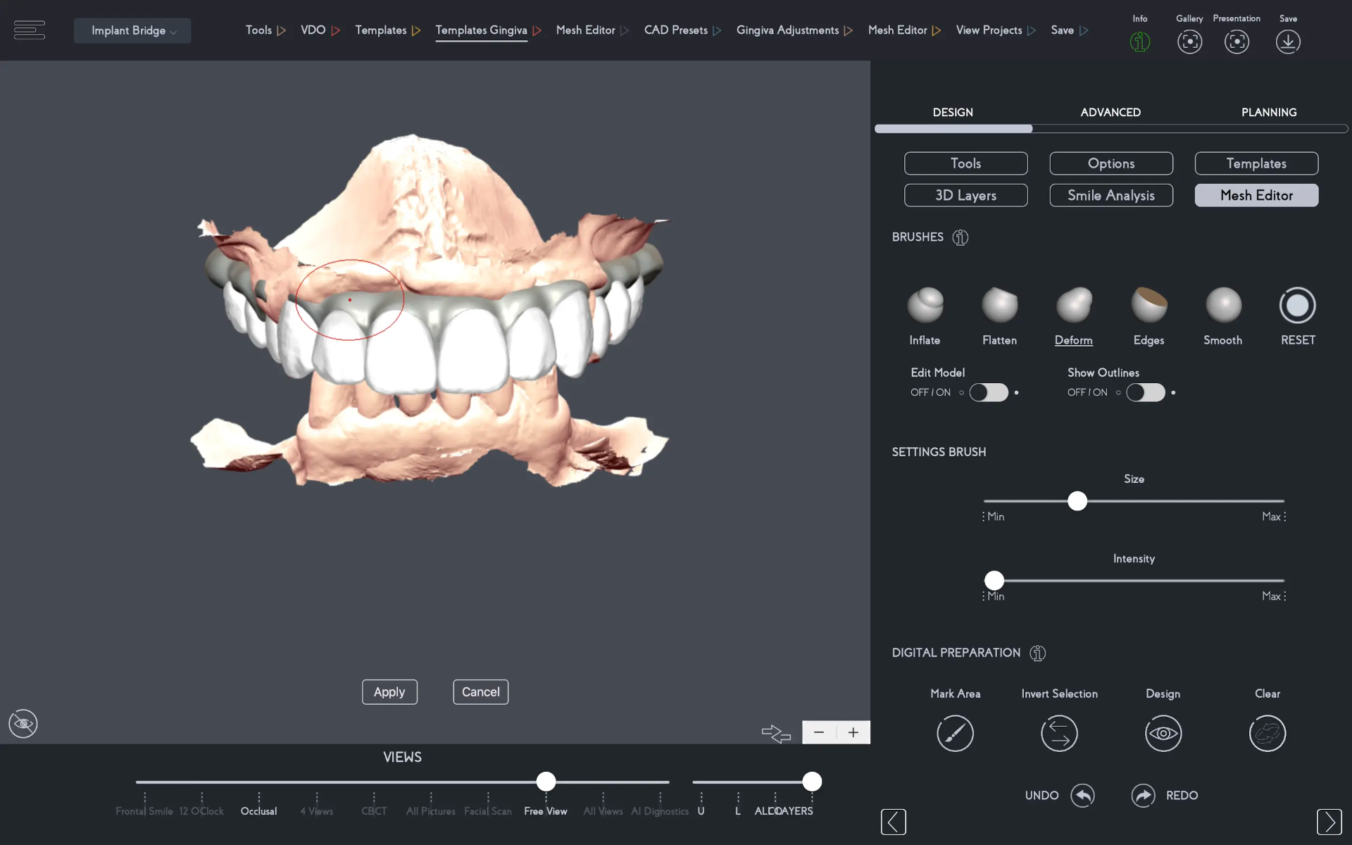
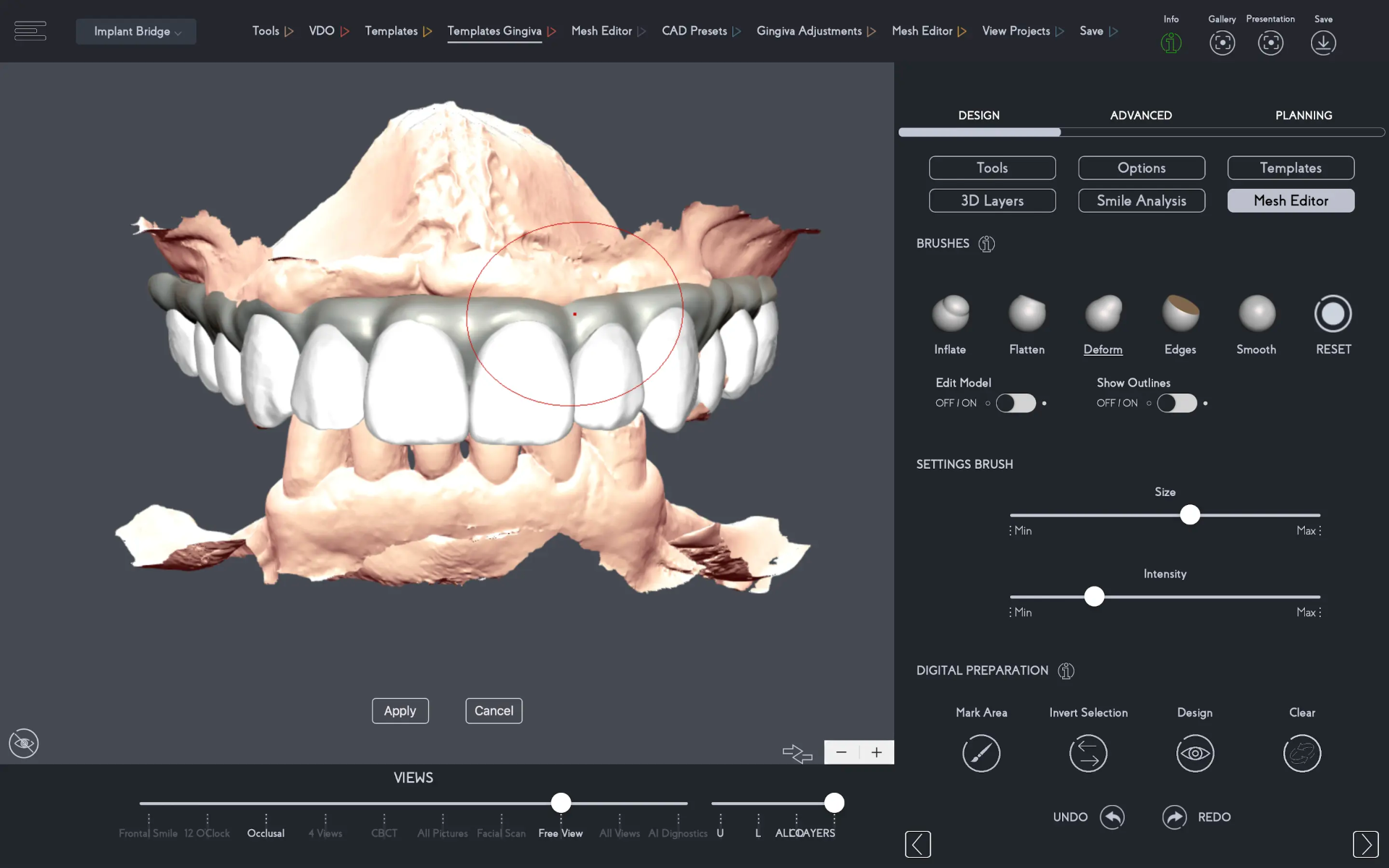
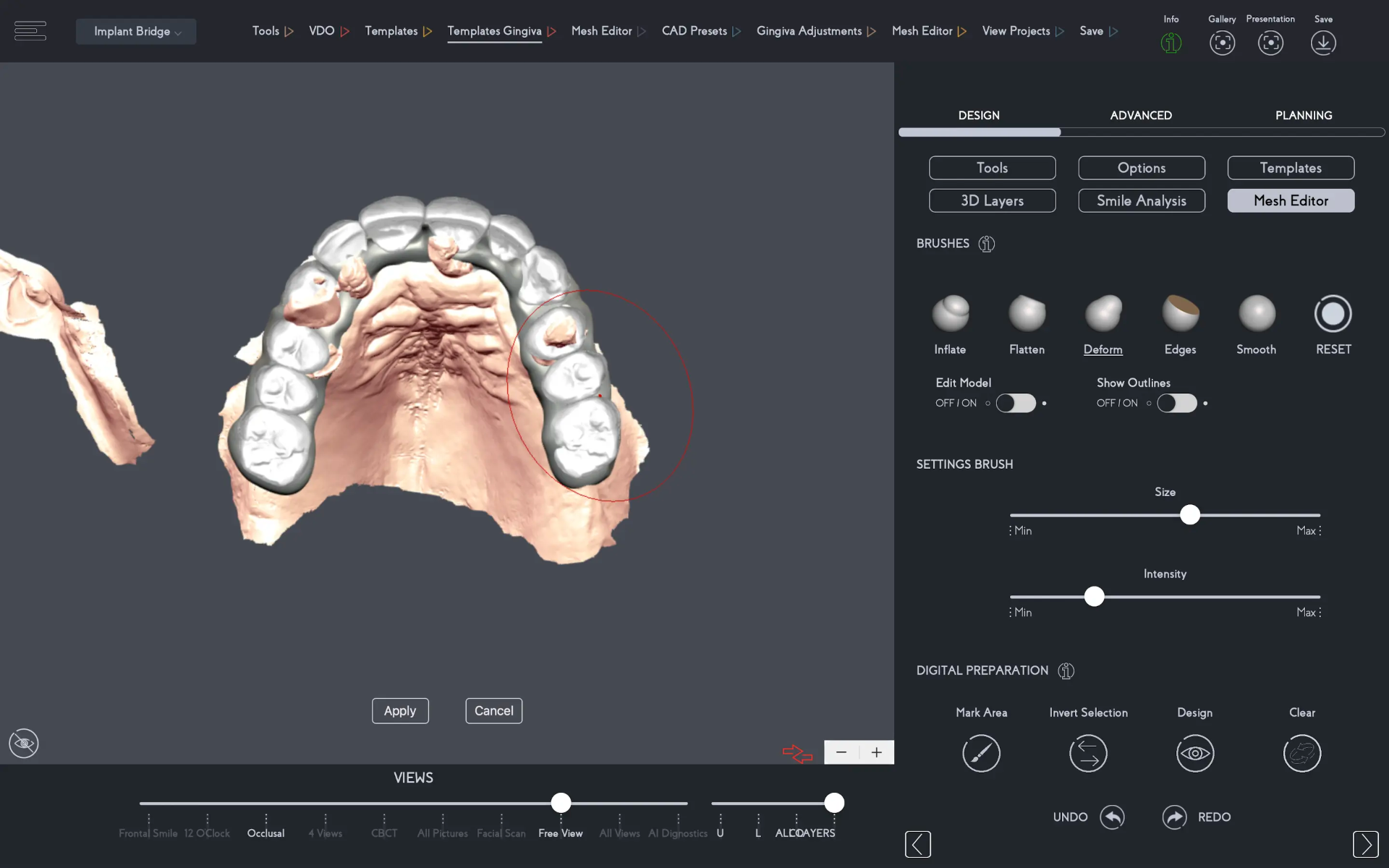
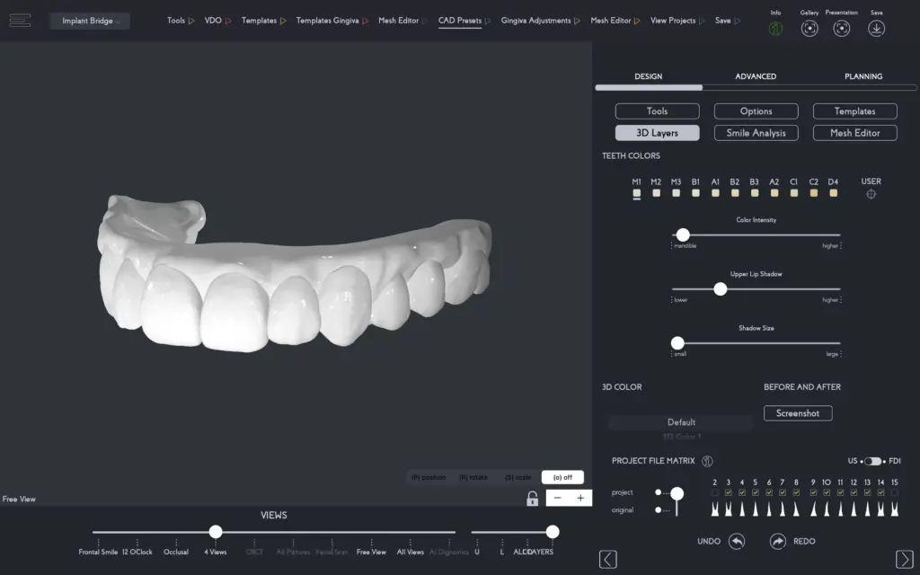
Before setting up the export of the implant bridge design, the dental professional has the option to add colors to the design to preview the case virtually first.
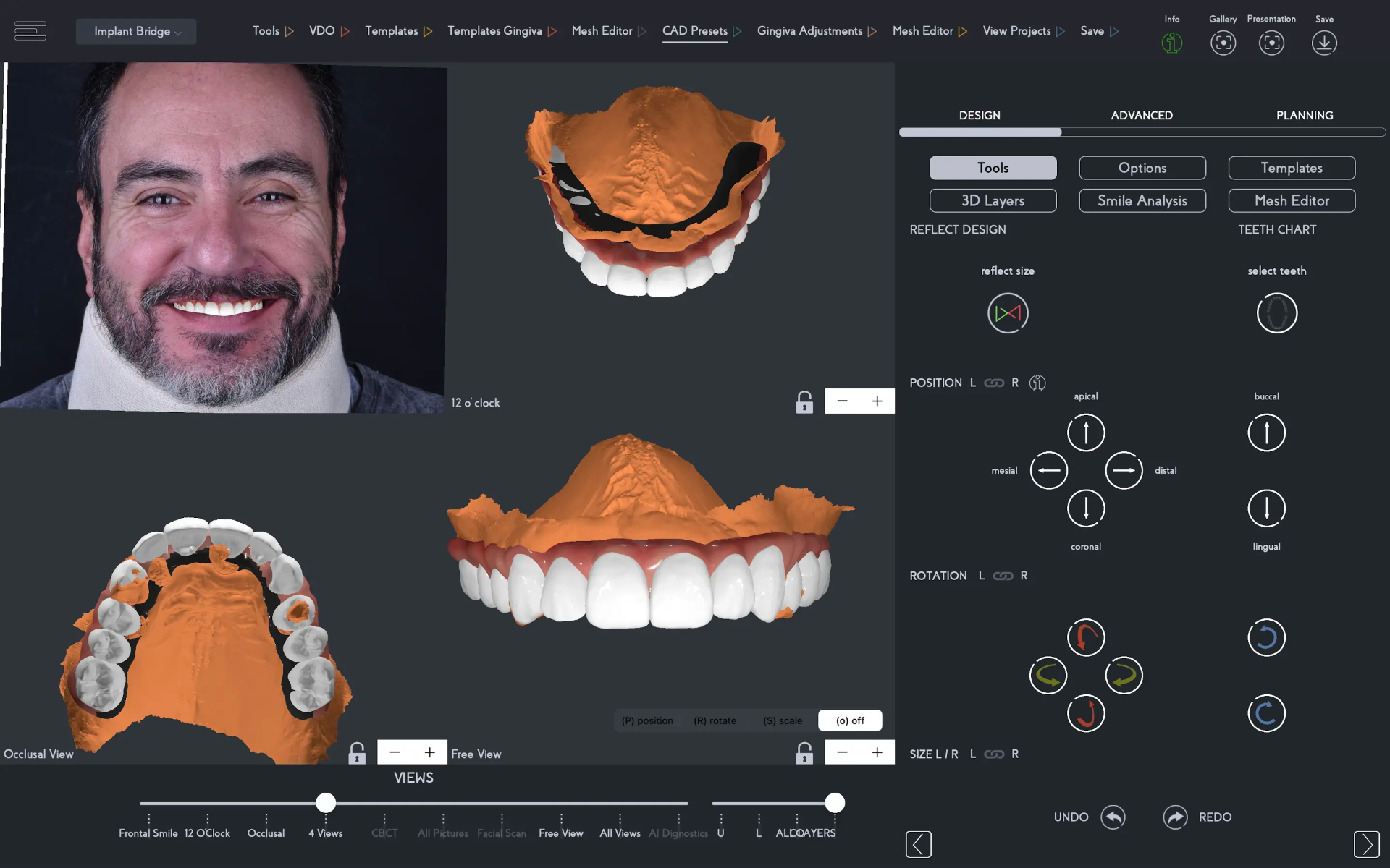
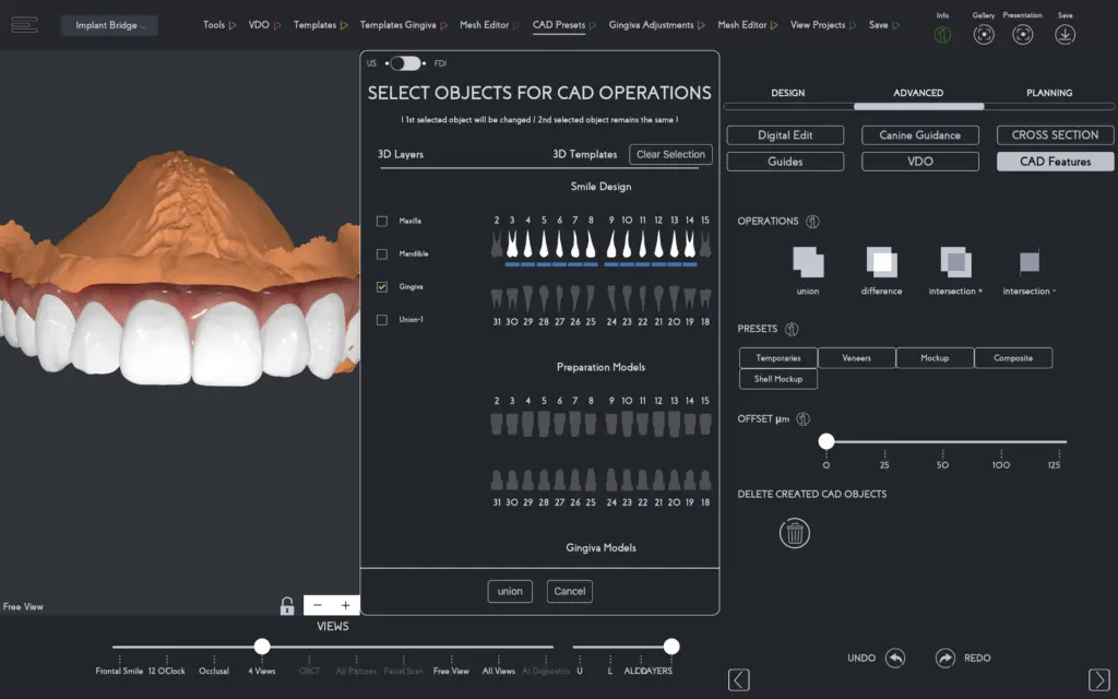
With the design ready, simply combine the 3D teeth and gingiva to create one solid exportable file.
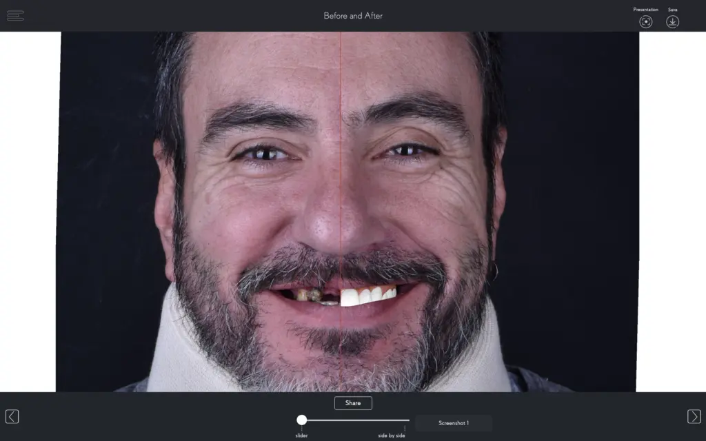
An important step is to send the patient and lab technician the before and after virtual implant bridge design for further visual reference of the treatment planning.
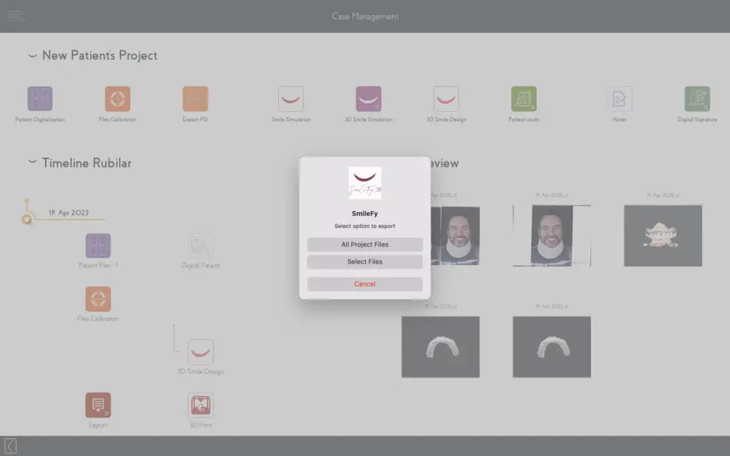
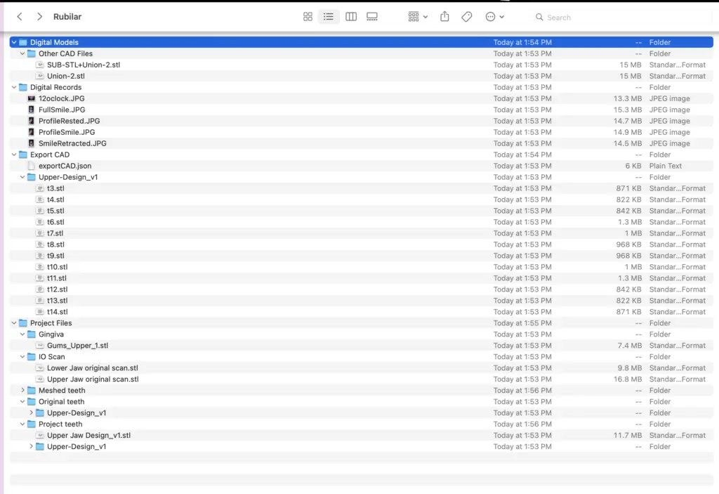
After saving the case, all files can be exported as .stl files, shared with any person, and opened in any other planning software that accepts .stl files. The files include the patient's original files untouched, as well as the 3D dental bridge design and the 3D teeth templates as separate files.
Using the 3D Bridge Design as Virtual Guidance for Implant Planning
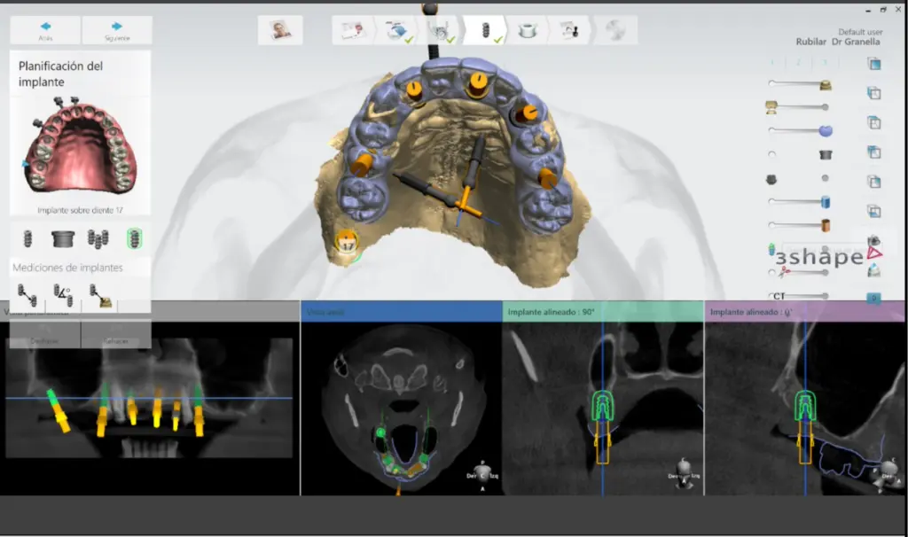
For the surgical planning steps, the exported 3D implant bridge design in .stl file format was added to an implant planning software, and a second superimposition of the files was performed: superimposing the virtual implant bridge design and the CBCT data allowing a prosthetically driven implant position planning.
From this planning, a surgical guide was designed and fabricated. Following the dentist's guidance, the temporary implant bridge design was finalized in the dental laboratory, then milled in PMMA, stained, finished, and glazed.
Surgical Day and Immediate Loading Implant Bridge Placement
The teeth were extracted, a flattening of the alveolar ridge was performed to create space for the prosthetic restoration, six implant placements were executed, and four implants on the lower posterior region.
A primary stability torque ranged from 30Ncm–40Ncm, enabling immediate loading and giving enough engaging strength to the implant-abutment connections via the fixation screw with 1mm to 1.5mm space left between the orthesis and the gum. The interim calcined cylinders were connected to the prostheses with acrylic resin, screw-retained with 10 Ncm torque, and the screw access holes were sealed with Teflon tape and composite resin, and post-operative management instructions were provided.
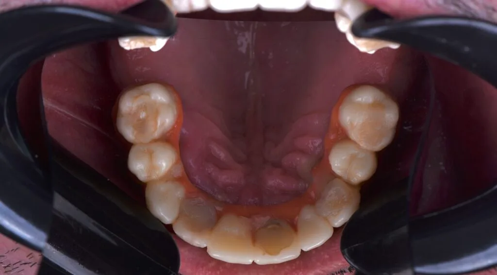
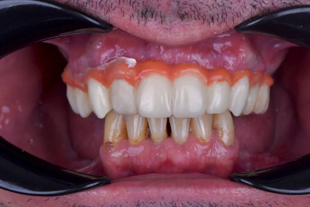
Five days later, a re-evaluation was made. The patient was very happy, without pain or inflammation.
After four to six months, the final crowns on the lower arch will be fabricated, and the upper fixed bridge will be fabricated based on the provisional designed on SmileFy.
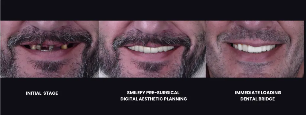
Conclusion
In the pre-surgical steps of extensive rehabilitation, the patient's face must be included in the treatment planning to help the clinician obtain positive esthetic and functional outcomes of the future prosthesis, which facilitates the digital planning of the surgical implant guides, and helps the lab technician to manufacture the intermediate and final prosthesis according to the clinician's plan.
SmileFy Planning software has been shown as an innovative process for Pre-Surgical Esthetic Planning, allowing dentists to superimpose the patient's photograph and digital casts to digitally pre-design the implant bridge while evaluating teeth contour, smile curvature, and proportions, enhancing the communication between the interdisciplinary team and patient towards a more incredible rehabilitative therapy and predictable outcome.
"What I love about the SmileFy workflow is that it gives me the autonomy to precisely pre-design my cases using a virtual patient, export it to an implant planning software and effectively guide the lab technician on fabricating the immediate loading provisional and final bridge." — Dr. Adrian Granella
Key Clinical Takeaways
Pre-surgical digital aesthetic planning — Essential for screw-retained prostheses to achieve facially guided, predictable outcomes
3D bridge design with SmileFy — Enables precise virtual planning of implant bridge contour, proportions, and gingival architecture before surgery
STL export workflow — Seamless file transfer to implant planning software for prosthetically driven implant positioning
Enhanced interdisciplinary communication — Visual documentation and 3D files bridge the gap between clinician, lab technician, and patient
Immediate loading protocol — Digital pre-planning supports confident same-day prosthesis delivery with predictable aesthetics
Video Walkthrough
Watch Dr. Adrian Granella's full video walkthrough of the digital dental bridge design process with SmileFy:
About the Author
Dr. Adrian Granella
SmileFy Member — G.Clinic, Mendoza, Argentina
Dr. Adrian Granella is a dental surgeon based at G.Clinic in Mendoza, Argentina. He specializes in extensive oral rehabilitation using digital workflows, with a particular focus on pre-surgical aesthetic planning for implant-supported prostheses. His clinical work demonstrates how SmileFy software can be integrated into complex implant cases to achieve predictable aesthetic and functional outcomes while streamlining communication between clinicians and dental technicians.
Ready to Transform Your Practice?
See how SmileFy can help you achieve predictable, beautiful results for every patient.