Digital Planning, Mockup Trials, and Interdisciplinary Management of Gummy Smile with Full-Arch Maxillary Rehabilitation
Dr. Elias Abou Tayeh
Aesthetic and Restorative Dentist — Diploma in Oral Biology, Implantology, TMJ & Occlusion
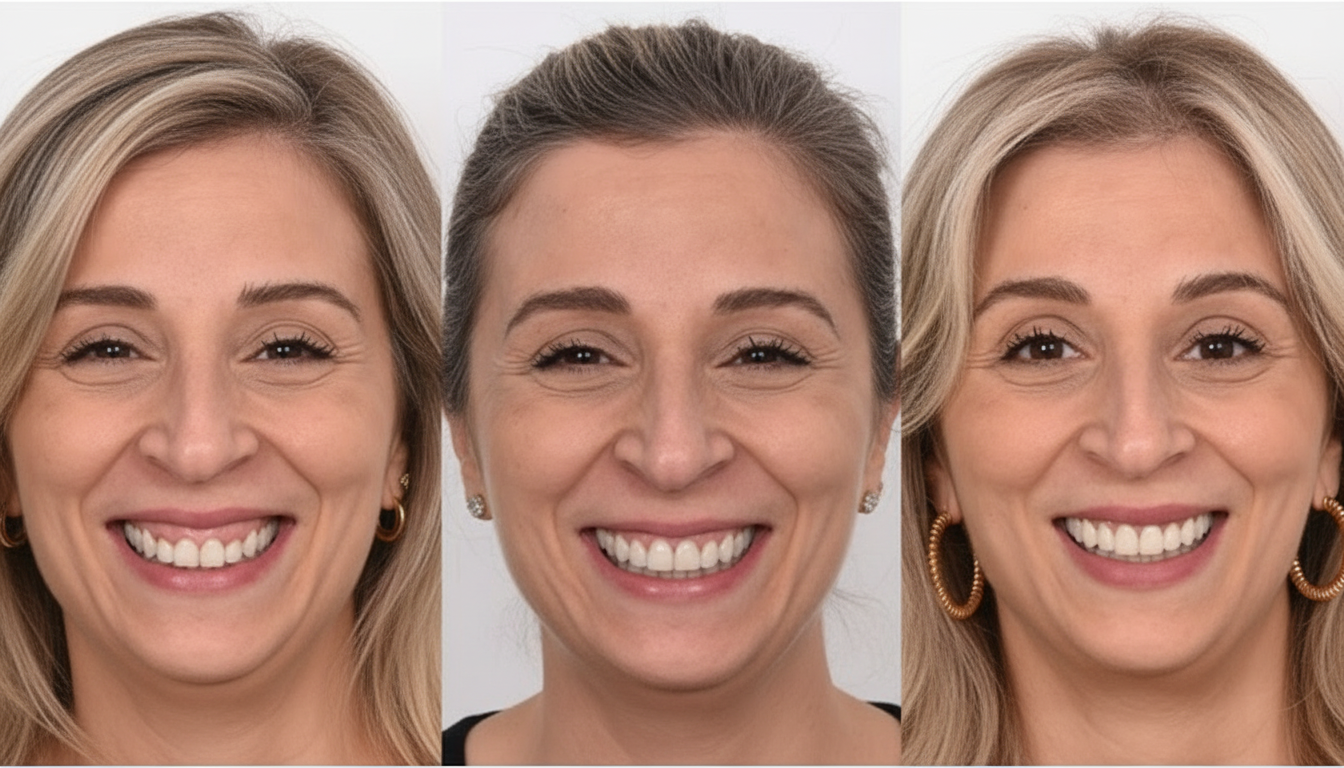
Patient Summary
Abstract
This case report describes the comprehensive aesthetic and functional rehabilitation of a 43-year-old female presenting with excessive gingival display and ageing ceramic crowns. Treatment was conducted through a fully integrated digital and interdisciplinary workflow incorporating AI-assisted 2D smile simulation (SmileFy), 3D-printed resin mockup fabrication, cone-beam computed tomography (CBCT)-guided surgical planning, crown lengthening with a digitally fabricated surgical guide, and delivery of full-arch maxillary ceramic restorations. The structured four-step protocol — from digital design through clinical execution — demonstrates how contemporary digital tools can improve outcome predictability, streamline interdisciplinary communication, and enhance the patient's informed consent experience.
Treatment Plan Overview
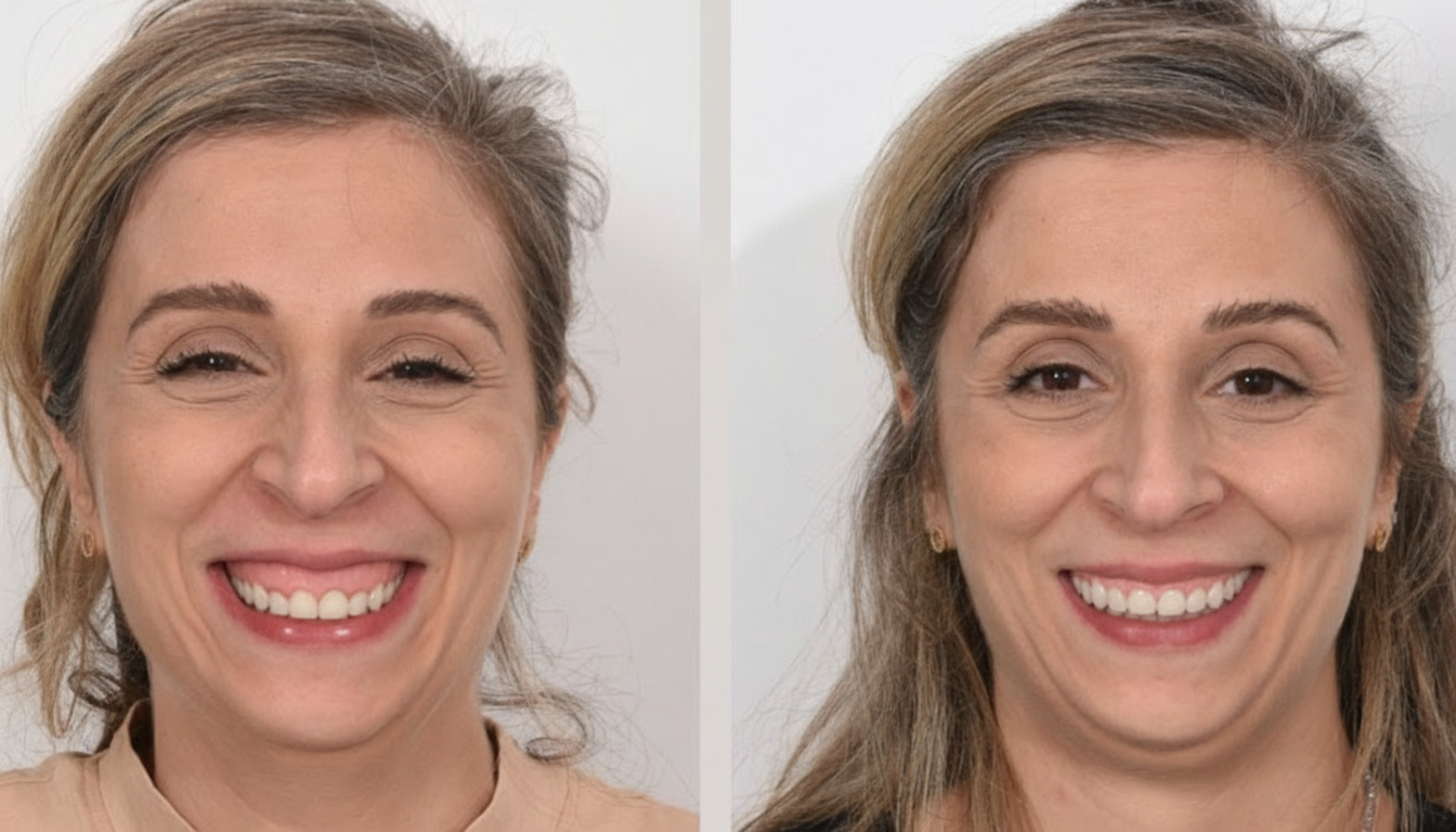
The primary objective was to harmonise the patient's smile by correcting a pronounced gingival display and replacing existing crowns with ceramic restorations proportioned and designed in accordance with a facially guided digital smile design. The plan aimed to deliver a balanced, natural, and aesthetically coherent result consistent with the patient's facial morphology. This was achieved through a multidisciplinary strategy uniting digital smile design, periodontal surgical correction, and definitive prosthodontic restoration. AI-driven planning tools informed all phases, maintaining a conservative, patient-centred approach that prioritised both function and aesthetics.
Interdisciplinary Scope
Three core disciplines were engaged in a coordinated workflow. Periodontics addressed gingival level correction and symmetry through crown lengthening. Prosthodontics was responsible for the design, fabrication, and delivery of the final ceramic restorations. Digital technology — specifically the SmileFy AI platform — provided the diagnostic and communication framework that aligned all disciplines throughout treatment, enabling precision design transfer from the digital simulation to the physical mockup and, ultimately, to the definitive restorations.
Workflow Summary
Step 1 — Diagnostic assessment and 2D digital smile design
Step 2 — In-clinic mockup trial (3D-printed resin): esthetics, speech, and occlusion checks
Step 3 — Interdisciplinary planning: CBCT analysis and surgical guide for crown lengthening
Step 4 — Final upper ceramic restorations: bonding and finishing
Step 1 — Diagnostic Assessment and 2D Digital Smile Design
Treatment commenced with a comprehensive facially guided diagnostic evaluation. The Facial Flow Concept was applied to systematically assess the interrelationship between the patient's facial structure, lip frame, smile arc, midline, and gingival display. Photographic documentation was obtained in standardised repose, posed smile, and retracted intraoral views.
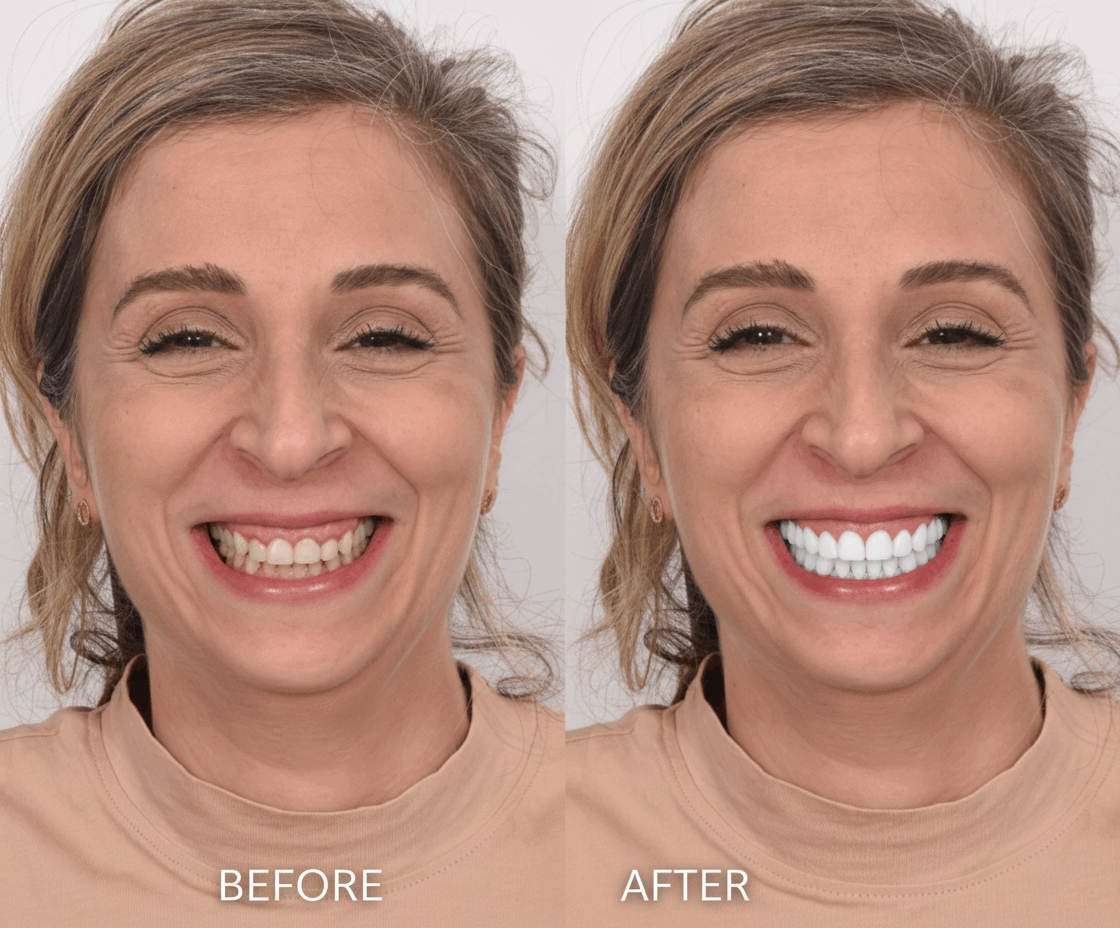
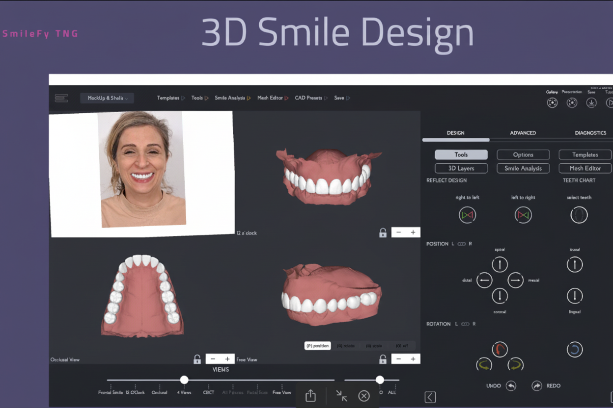
SmileFy AI software was employed to generate a 2D digital smile simulation. This visualisation allowed the clinical team to establish tooth proportion targets, define the proposed gingival zenith positions, and map the ideal incisal edge positions relative to the lower lip curvature. The resulting digital design served as a blueprint for all subsequent clinical and laboratory phases. Critically, the simulation also functioned as a structured communication tool. Presenting the anticipated outcome to the patient at this early stage promoted a shared understanding of treatment objectives, facilitated informed consent discussions, and allowed expectation calibration before any clinical procedures were undertaken.
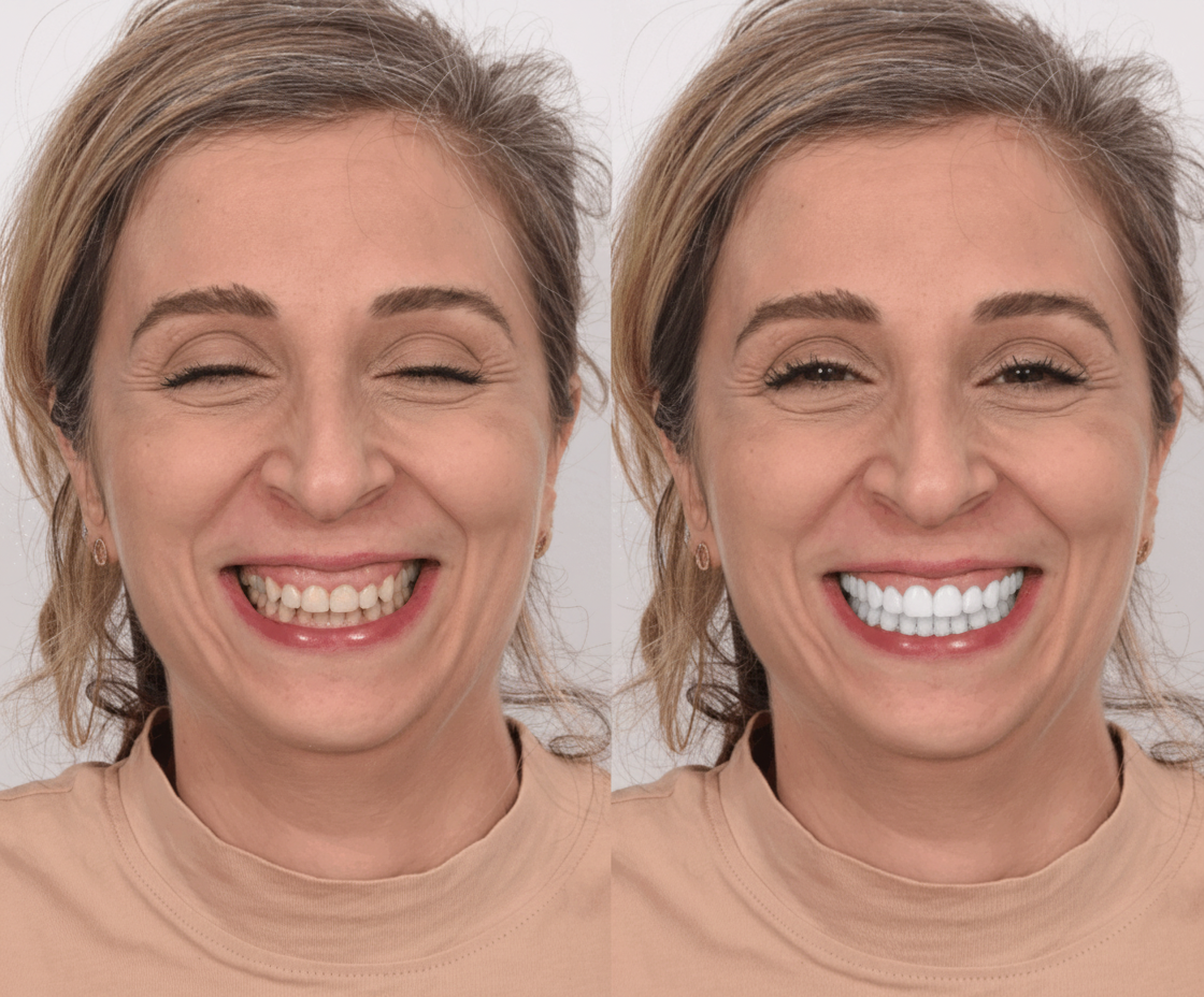
Step 2 — In-Clinic Mockup Trial (3D-Printed Resin): Esthetics, Speech, and Occlusion Checks
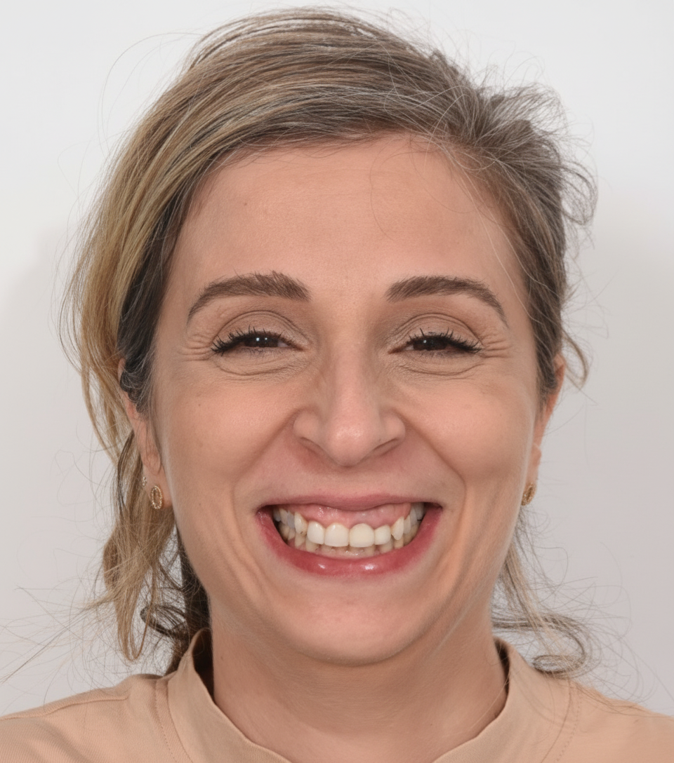
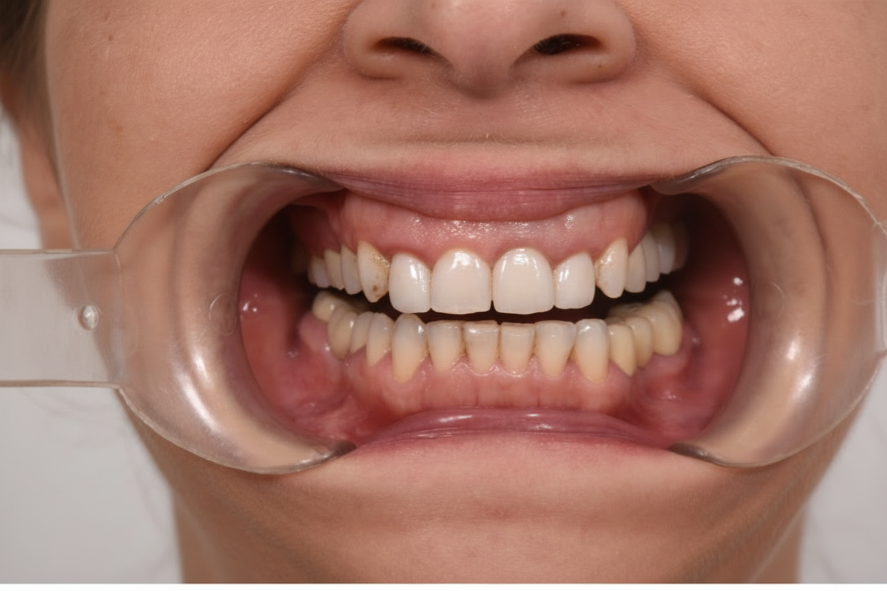
Once the 2D digital design was approved by both the patient and the clinical team, a physical mockup was fabricated from 3D-printed resin materials. This in-clinic trial served as a tangible, real-time prototype of the planned aesthetic outcome, enabling evaluation dimensions not accessible through digital visualisation alone.
The mockup was seated intraorally and assessed across three critical parameters:
Esthetics — Viewed under clinical and natural lighting from multiple angles; patient and clinician feedback documented.
Speech — Phonemic assessment of sibilants (particularly /s/ and /f/ sounds) to detect potential occlusal or incisal edge position interference.
Occlusion — Preliminary occlusal contacts and dynamic movements evaluated for premature contacts or lateral interferences introduced by the proposed tooth dimensions.
The mockup session allowed iterative refinements to be made to tooth length, width, and surface texture before any irreversible clinical procedures were initiated. Patient responses and reactions were formally documented, and minor design modifications were incorporated into the final digital design file prior to progression to the interdisciplinary planning phase.
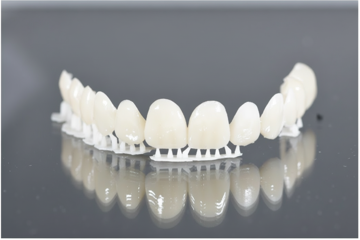
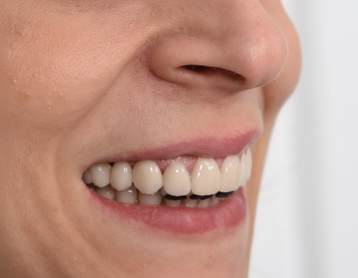
Step 3 — Interdisciplinary Planning: CBCT Analysis and Surgical Guide for Crown Lengthening
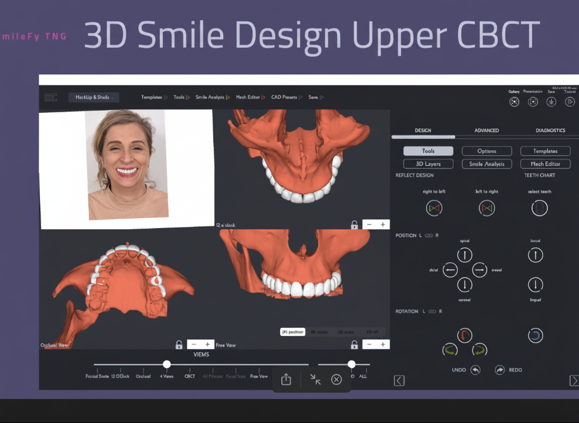
Following mockup approval, the clinical team proceeded with detailed surgical and restorative planning. CBCT imaging was obtained and analysed to assess bone levels, tooth root morphology, crestal-to-CEJ distances, and available restorative space. Diagnostic wax-ups were prepared and cross-referenced with the CBCT data to quantify the degree of hard and soft tissue recontouring required to achieve the planned gingival margin positions.
A 3D-printed surgical guide was fabricated from the digital smile design to precisely direct the extent of gingival and osseous recontouring during the crown lengthening procedure. This guide communicated the target gingival margin positions — as established in the digital simulation and confirmed in the mockup trial — directly to the operating periodontist, eliminating reliance on subjective intraoperative estimation.
The periodontist performed gingival recontouring and, where indicated by crestal bone proximity, osseous correction to establish adequate biologic width clearance. The surgical guide ensured that tissue removal was conservative, predictable, and consistent with the pre-approved digital design. A structured healing period was observed post-surgery to allow soft tissue maturation before proceeding to the restorative phase.
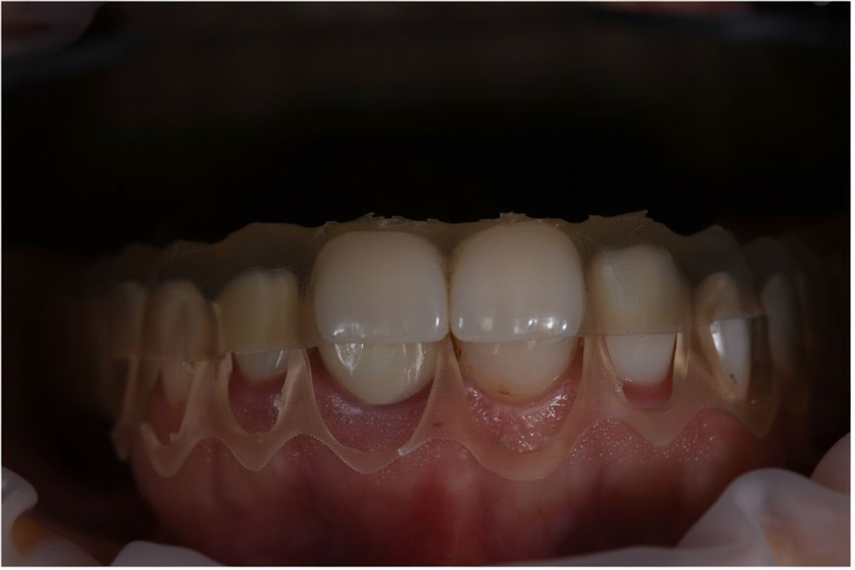
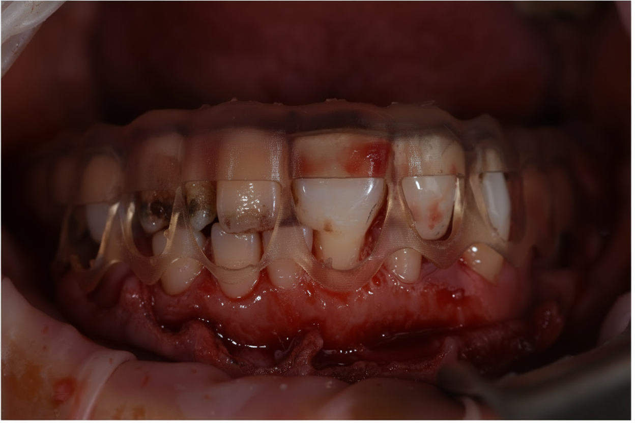
Step 4 — Final Upper Ceramic Restorations: Bonding and Finishing
Following soft tissue healing and confirmation of stable gingival margins, the final restorative phase commenced. Tooth preparations were completed in accordance with the pre-planned digital design parameters, respecting the newly established gingival architecture.
Definitive ceramic restorations were designed using the SmileFy digital smile design files, ensuring precise dimensional continuity from the approved mockup to the laboratory-fabricated crowns. The restorations were prescribed to replicate the mockup in tooth form, length, and shade, with the advantage that the patient had already previewed and approved the aesthetic result in situ.
The bonding procedure was carried out with meticulous attention to marginal adaptation and seating accuracy. Occlusal contacts were refined in both intercuspation and excursive movements. Final polishing was performed to achieve optimal surface lustre and a natural, vibrant aesthetic. The completed restoration was assessed for occlusal function, soft tissue health at the margins, and aesthetic integration with the facial frame.
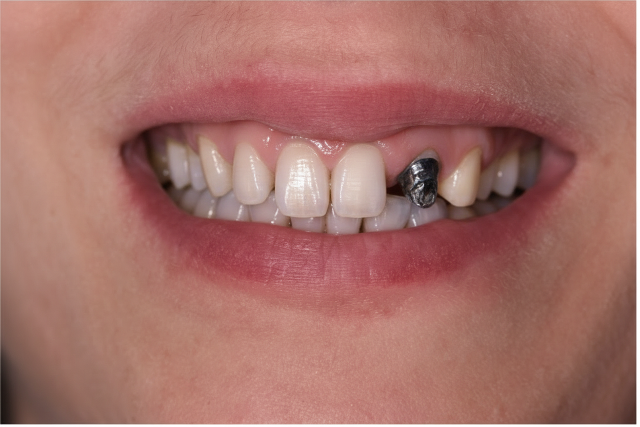
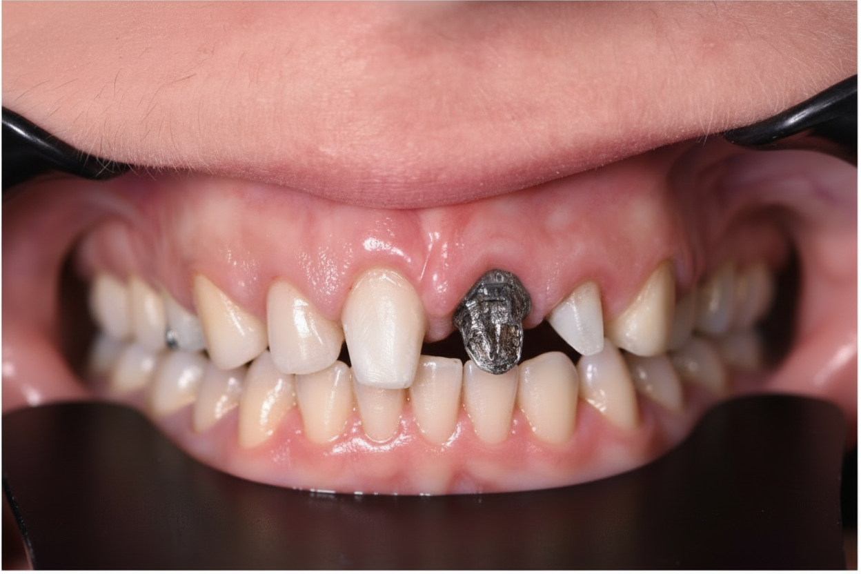
Final Outcome
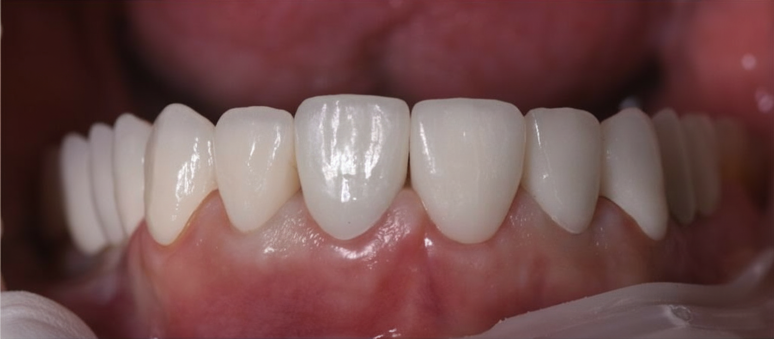
The treatment resulted in a clinically significant transformation in the patient's smile aesthetics and oral harmony. Excessive gingival display was successfully corrected, and the maxillary teeth were restored to proportions consistent with the digital design targets. The final ceramic restorations demonstrated excellent marginal integrity, shade matching, and occlusal function.
Post-treatment photographs confirmed harmonious integration between the restored teeth, the corrected gingival contour, and the patient's broader facial aesthetics. The patient expressed high levels of satisfaction with both the process and the outcome. Comprehensive oral hygiene instructions were provided alongside a structured recall protocol to support the longevity of the periodontal and prosthetic results.
Case Reflection and Clinical Insights
The principal clinical challenge in this case was achieving an appropriate balance between soft tissue recontouring and the creation of adequate restorative space. Maintaining the biologic width during gingival correction was paramount to prevent post-operative complications, including recurrent inflammation, marginal recession, or prosthetic margin exposure. This was addressed through CBCT-guided pre-surgical planning and the use of the digitally fabricated surgical guide, which together enabled precise, controlled osseous recontouring.
A secondary challenge was ensuring dimensional and aesthetic fidelity between the approved digital mockup and the definitive laboratory restorations. This required close coordination between the designing clinician and the ceramist, with the SmileFy digital files serving as the shared reference throughout the fabrication process.
Role of Digital Technology
The SmileFy AI platform provided a cohesive digital thread linking all phases of treatment. The 2D smile simulation established the design target; the 3D-printed mockup made that target tangible and testable; and the digital files transferred the approved design to both the surgical guide and the laboratory with precision. This workflow reduced interpretive ambiguity between disciplines and significantly improved outcome predictability.
The ability to simulate, evaluate, and modify the proposed outcome prior to any irreversible intervention also proved invaluable for patient communication. Patients who can visualise and physically trial their future smile prior to preparation are better equipped to make informed decisions and demonstrate higher satisfaction throughout the treatment process.
Key Clinical Takeaways
Digital smile design using AI-assisted tools — Enables precise, reproducible aesthetic planning and provides a shared reference for all members of the interdisciplinary team.
3D-printed resin mockups — Constitute a critical pre-treatment validation step, allowing simultaneous assessment of aesthetics, phonetics, and occlusal compatibility before any irreversible procedures are performed.
Differential diagnosis of gummy smile aetiology — Essential prior to selecting crown lengthening; altered passive eruption, vertical maxillary excess, hyperactive lip, and dentoalveolar extrusion require distinct management strategies.
Biologic width preservation — Must be explicitly planned in the surgical design; CBCT-guided crown lengthening with a digitally fabricated guide supports predictable, conservative osseous correction.
Dimensional continuity — From digital design through mockup to definitive restoration requires close clinician–ceramist collaboration and a robust digital transfer protocol.
Integration of digital innovation with disciplined clinical execution — Produces outcomes that are predictable, patient-centred, and durable — establishing a replicable model for complex aesthetic-functional rehabilitation cases.
About the Author
Dr. Elias Abou Tayeh
Aesthetic and Restorative Dentist — Diploma in Oral Biology, Implantology, TMJ & Occlusion
Dr. Elias Abou Tayeh is an aesthetic and restorative dentist based in Beirut, Lebanon, with advanced training in oral biology, implantology, TMJ, and occlusion. He is the founder and CEO of Fixaligner, a company combining digital dentistry technology with affordable clear aligner solutions. His clinical work focuses on integrating AI-driven digital smile design with interdisciplinary treatment planning.
Ready to Transform Your Practice?
See how SmileFy can help you achieve predictable, beautiful results for every patient.