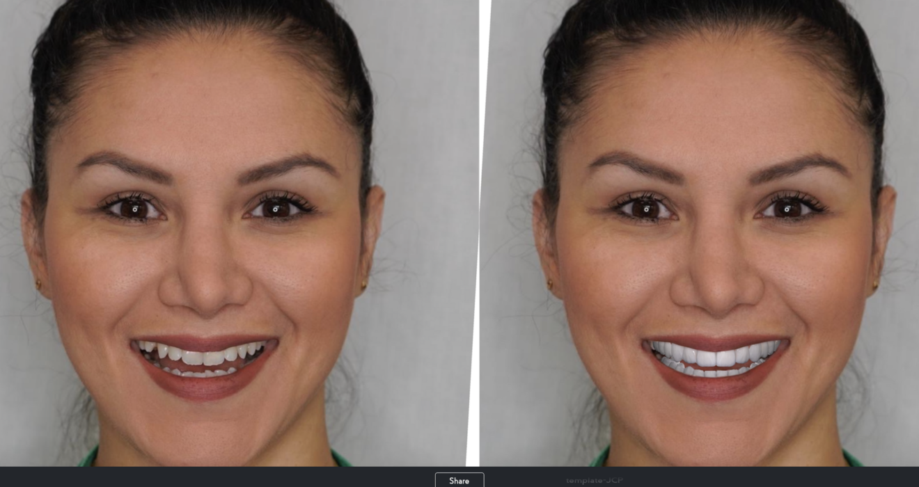
3D A.I. Smile Simulation: How to Perform Smile Harmony in Asymmetrical Faces
Author: Dr. Diogo Alves
Deep studying your patient’s facial anatomy helps you to achieve higher quality dental esthetic outcomes in cosmetic dentistry, But do you know where to start?
In this blog, you will learn how to perform Smile Study Analysis using the dentist-friendly SmileFy Software in:
- Proper Picture Protocol
- A.I. Auto-Capture
- Principles of Smile Design Analysis
- A.I. Smile-Facial Analyzer in the 3D Smile Simulator
As clinicians, we must pay attention to delivering the function and how to customize the position, angulation, and shape of the teeth according to the characteristics of the patient’s face. Severt and Proffit reported frequencies of facial laterality of 5%, 36%, and 74% in the upper, middle, and lower thirds of the face, respectively, with a more significant variation in the lower third of the face [3]. So let’s dive into how simple in-office digital methods can help us deliver a more aesthetically pleasing, facially driven smile.
“To Achieve a successful, healthy, and functional result requires you to understand the interrelationship among all the supporting oral structures, combining the muscles, bones, joints, gingival tissues, and occlusion.”[2] . Article: Dawson PE. Determining the determinants of occlusion. Int J Periodontics Restorative Dent. 1983;3:8–21.
The Photograph
Among the various photographs we took in our field, such as intraoral photos, profile photos, and 45 degrees, let’s discuss the frontal image with an open smile to start our orofacial aesthetic analysis.A photograph of a spontaneous smile must be captured to show the maximum elevation of the upper lip with a spontaneous smile and the mouth slightly opened to evaluate the parallelism of the curvature of the upper arch with the lower lip. helping to identify deviations of the upper and lower occlusal planes compared to the face. [5]
SmileFy A.I. Auto-Capture
To help dental professionals better photograph their patients, SmileFy created the “A.I Auto-Capture,” a photograph tool that uses Artificial Intelligence technology to help you take better pictures through the automated detection of a proper eye, head, and smile position for the smile design.
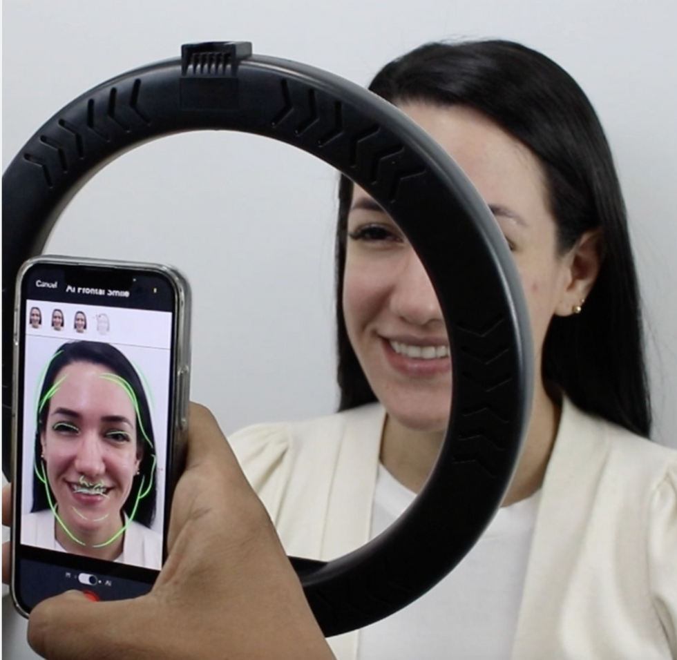
Position your patient straight up, with no hats or glasses and hair tucked behind the ears. In a room with great lighting, frame the patient’s face inside the screen of your camera, eliminating shoulders.
To prevent distortions on the smile curve of the patient’s current conditions, level the camera’s lens to your patient’s eyes and ensure that your camera is straight. Looking at the camera’s lens, ask your patient to give a wide smile with their mouth slightly open for the picture to be taken with a.i. technology.
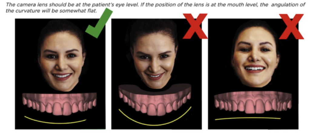
Principles of Smile Design Analysis
There are three facial features which do play a major role in the smile design from a frontal facial view:
- The interpupillary line
- Lips
- Facial Midline
And three dental features also play an important role in the smile design from a frontal facial view:
- Position of central incisors
- Smile Curvature
- Position of the occlusal plane
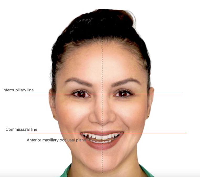
SmileFy A.I. Smile-Facial Analyser
Using SmileFy Software, you are able to analyze these lines using your patient’s image, which will help you define the position and shape of the future treatment and restorations, giving a boost of confidence to your treatment goals and ultimately enhancing the communication with your lab and patients
- The interpupillary line should be perpendicular to the midline of the face and parallel to the occlusal plane from an anterior view
- The incisal edge position of the central incisors should be perpendicular to the interpupillary line
- The smile curvature should follow the shape of the lower lip line
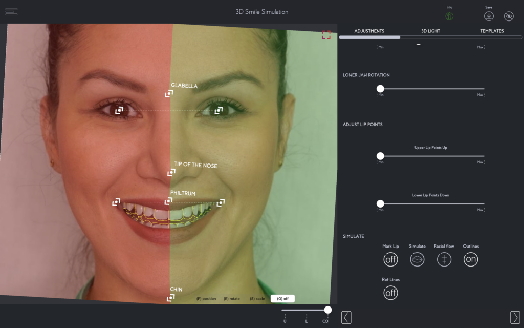
And the position of the central incisors should follow the imaginary facial line consisting of the glabella’s center, the nose tip, the philtrum ( cupid bow), and the chin tip.
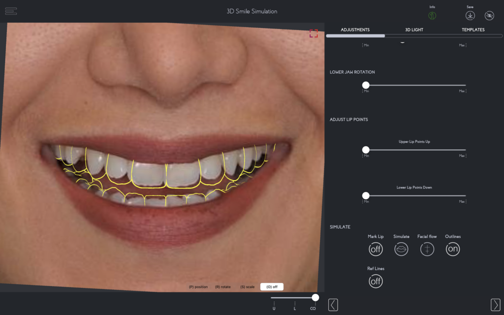
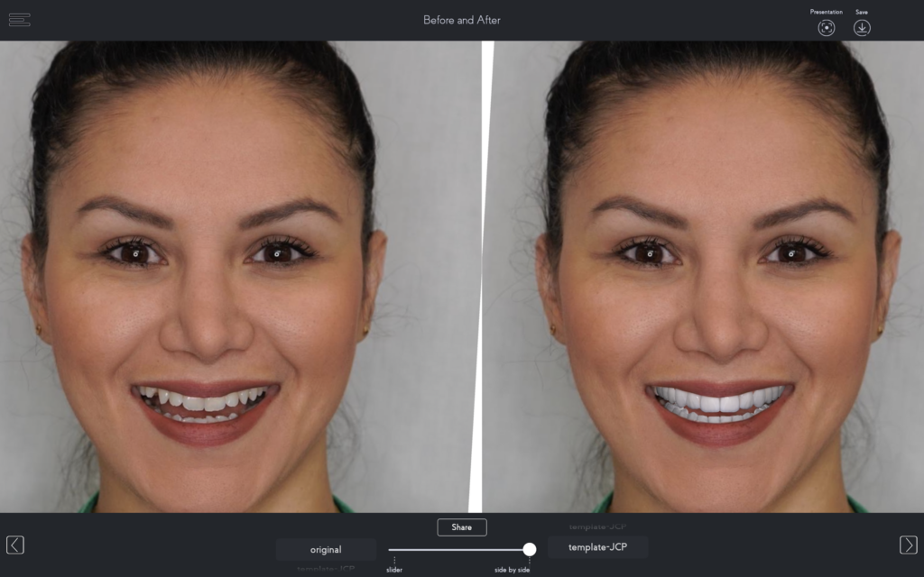
As part of your analysis and documentation, SmileFy offers 3D natural teeth that you can select from a variety of templates to be virtually tested in seconds and used as a reference further to enhance the communication with your patient and lab as well as to continue the planning into a 3D Smile Design ans a Print-ready Mockup file
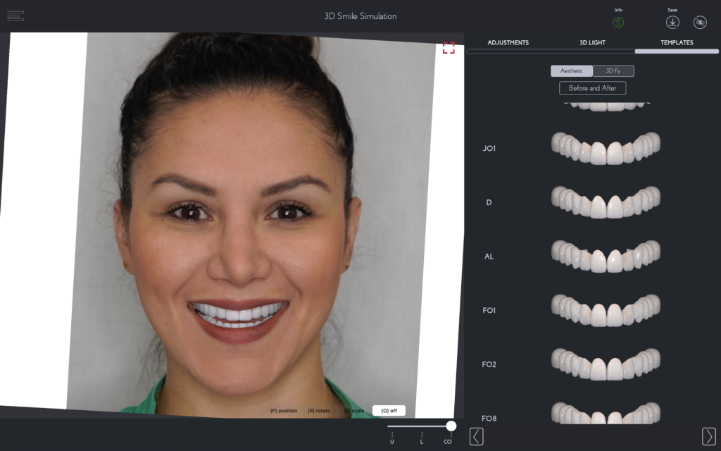
For a more comprehensive treatment plan, using your clinical knowledge, SmileFy Smile Design can be applied to digital visual planning, giving you better aesthetic knowledge and the ideal position of teeth in the upper arch in relation to the face for each patient, facilitating communication with other dentists and involving them in the multidisciplinary treatment workflow.
References:
1-Thiesen G, Gribel BF, Freitas MP. Facial asymmetry: a current review. Dental Press J Orthod. 2015;20(6):110-125. doi:10.1590/2177-6709.20.6.110-125.sar https://www.ncbi.nlm.nih.gov/pmc/articles/PMC4686752/
2 – Dawson PE. Determining the determinants of occlusion. Int J Periodontics Restorative Dent. 1983;3(6):8-21. PMID: 6584415. https://pubmed.ncbi.nlm.nih.gov/6584415/
3 – Severt TR, Proffit WR. The prevalence of facial asymmetry in the dentofacial deformities population at the University of North Carolina. Int J Adult Orthodon Orthognath Surg. 1997;12(3):171-6. PMID: 9511487. https://pubmed.ncbi.nlm.nih.gov/9511487/
5 -Jayalakshmi NS, Ravindra S, Nagaraj KR, Rupesh PL, Harshavardhan MP. Acceptable Deviation between Facial and Dental Midlines in Dentate Population. J Indian Prosthodont Soc. 2013;13(4):473-477. doi:10.1007/s13191-012-0234-6 https://www.ncbi.nlm.nih.gov/pmc/articles/PMC3792300/5 – Padwa BL, Kaiser MO, Kaban LB. Occlusal cant in the frontal plane as a reflection of facial asymmetry. J Oral Maxillofac Surg. 1997 Aug;55(8):811-6; discussion 817. doi: 10.1016/s0278-2391(97)90338-4. PMID: 9251608. https://pubmed.ncbi.nlm.nih.gov/9251608/

