Smile Design in restorative dentistry has helped clinicians achieve optimal results for their patients while simultaneously improving their treatment planning skills.
That being said, the three types of scenarios we most commonly hear from dentists when it comes to Digital Smile Planning narrows down to:
- Dentists who hear about smile designs but find it challenging to implement this workflow in their practice because they assume they don’t have time to apply it in their day-to-day
- Dentists who benefit from using technology by employing both analog and digital workflows for more predictable dentistry focused on multidisciplinary and conservative rehabilitation
- Dentists who are too comfortable with analog workflows and are hesitant at trying something new or outside of their comfort zone
Whichever type of dentist you can associate yourself with, digitally planning your smiles helps improve your aesthetic clinical planning and gives the patient a better understanding of the treatment that you propose.
Before starting your journey in the world of Digital Smile Planning, it is necessary to first understand why you are carrying out visual planning before starting the treatment, which is often irreversible.
Patient Centricity
The primary path is to understand why your patient came to your clinic and know how you can help them have oral health that goes beyond their expectations.
Why?
What our patients see is not always the only problem to be solved to ensure the quality of health.
How?
Through clinical exams, photographs, and supplementary imaging examination, we are able to identify the path to why more focused attention on multidisciplinary dentistry will establish more comfort and longevity in the aesthetic and masticatory balance.
When?
Primary care should be done at your first appointment. When you receive your patient, your eyes shouldn’t be focused only on the oral cavity but also on the patient’s face, paying close attention to the way the patients speak and the movements of their mouth and lips.
Your patient’s face provides crucial information for creating a balanced treatment.
Most of us have faces that are not symmetrical with the patterns given from scientific articles. This is due to proper genetic functions, posture, or habits. Thus, focusing only on the oral cavity can lead to a mismatch in harmony between the smile and the face and future occlusal balance due to lack of attention on the face. See the example below:
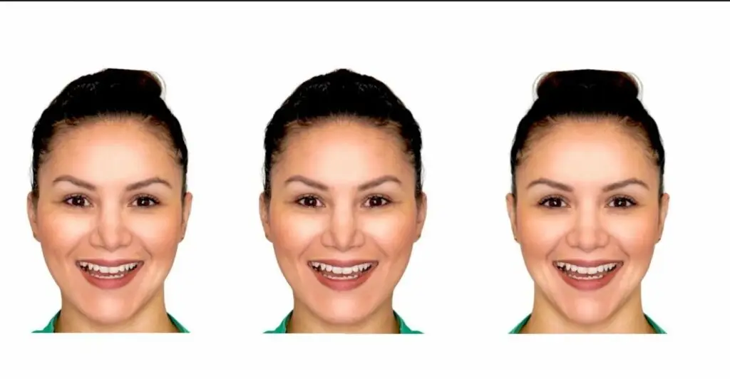
[Figure 1] A frontal facial smiling picture of a patient was taken. With its respective mirror image two other pictures were created: both right sides and both left sides showing that assessment of these images evinces the existing bilateral facial discrepancies.
Severt and Proffit reported frequencies of facial laterality of 5%, 36% and 74% in the upper, middle and lower thirds of the face respectively. [3] With a more significant variation in the lower third of the face, we as clinicians must pay attention on how to deliver our work where not only the function is provided, but also how to customize the position, angulation, and shape of the teeth according to the characteristics of the patient’s face.
Suppose we are guided by the aesthetic standards given by the media, evaluating and treatment planning. In that case, facial/dental esthetics often involves a multidisciplinary approach, including orthodontics, orthognathic surgery, periodontal therapy, cosmetic dentistry, and plastic surgery. However, in our clinical day-to-day, as long as the facial discrepancies are within the acceptable standard [4], the dentist should extol the patient’s beauty and plan a smile that will match the uniqueness of his/her face.
Finding the the right angle will help you determine:
Your patients facial midline
Your patient’s smile curvature
There are three facial features which do play a major role in the smile design:
The interpupillary line
Lips
Facial Midline
And three dental features which also play an important role in the smile design:
Position of central incisors
Smile Curvature
Position of the occlusal plane
Where?
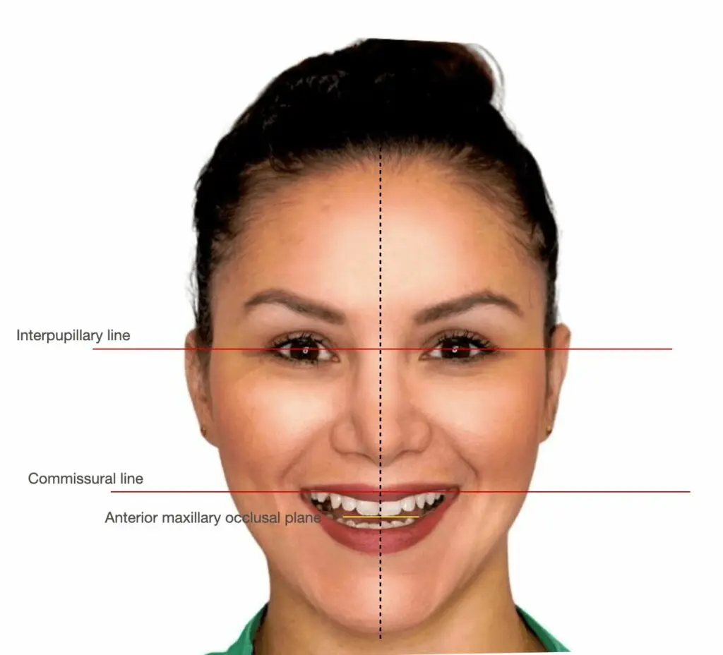
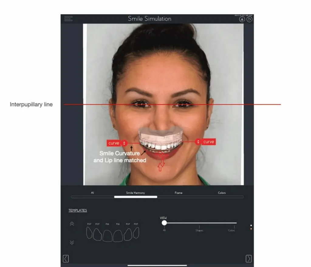
[Figure 2] The interpupillary line should be perpendicular to the midline of the face and parallel to the occlusal plane from an anterior view.
[Figure 3]
The incisal edge position of the central incisors should be perpendicular to the interpupillary line
The smile curvature should follow the shape of the lower lip line
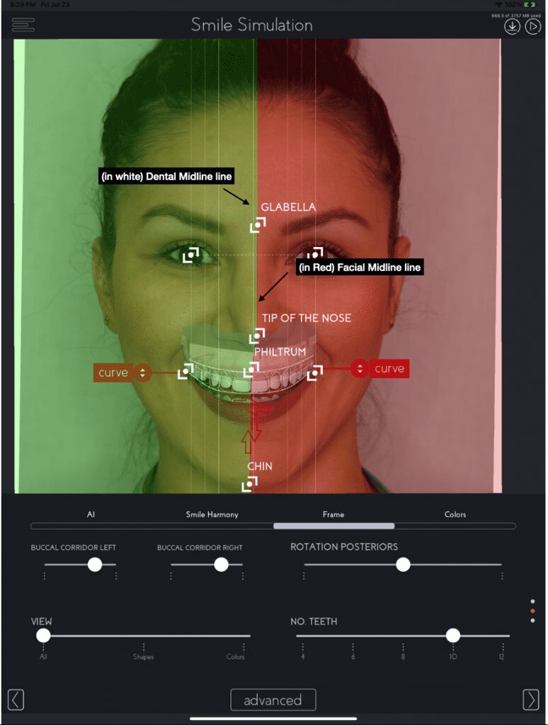
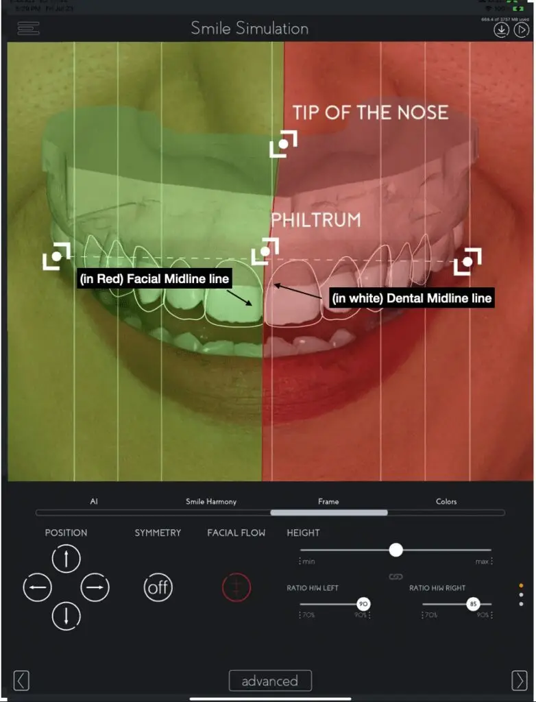
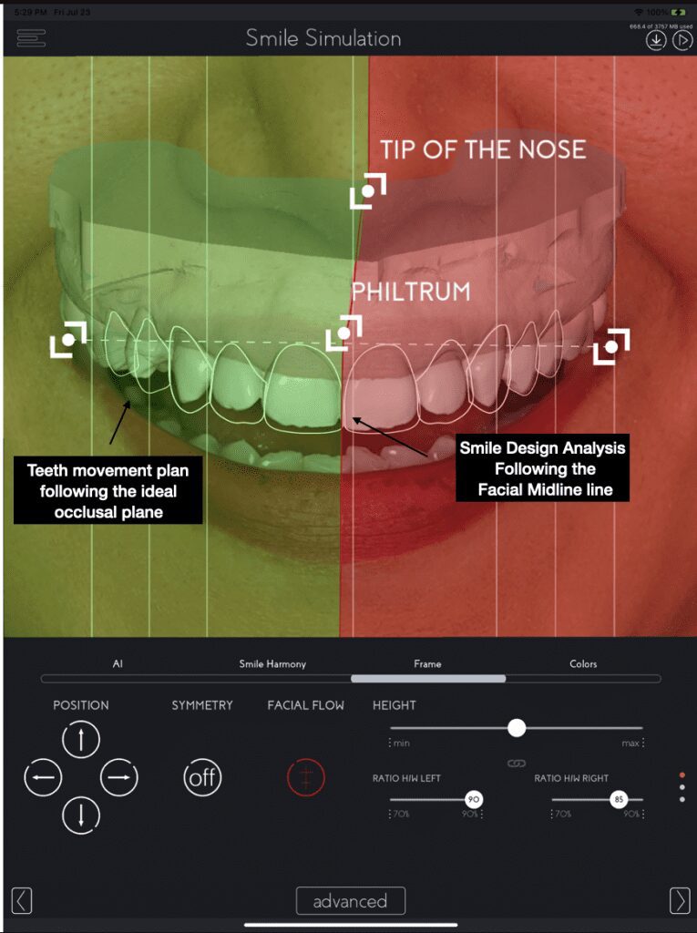
[Figures 4-6] And the position of the central incisors should follow the facial imaginary line that consists of the center of glabella, tip of the nose, philtrum (cupid bow) and tip of the chin.
Photographs bring to life imperfections not readily visible to the patient – and sometimes, you.
To make these observations, a good photograph of the patient must be taken.
Among the various photographs we took in our field, such as intraoral photos, profile photos, 45 degrees, let’s talk about the frontal image with an open smile to start our orofacial aesthetic analysis.
A photograph of a spontaneous smile must be captured to show the maximum elevation of the upper lip with a spontaneous smile and the mouth slightly opened, to evaluate the parallelism of the curvature of the upper arch with the lower lip helping to identify deviations of the upper and lower occlusal planes in comparison to the face. [5]
Position your patient standing up straight, with no hats or glasses, and hair tucked behind the ears. In a room with great lighting, frame the patient’s face inside the screen of your camera eliminating shoulders.
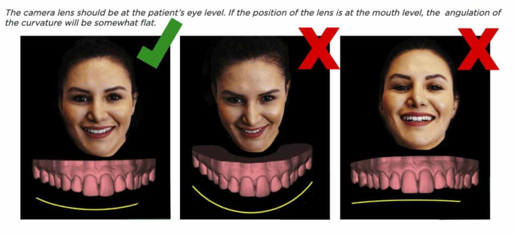
[Figure 7] To prevent distortions on the smile curve of the patient’s current conditions, level the lens of the camera to the eyes of your patient and make sure that your camera is straight. Looking at the lens of the camera, ask your patient to give a big wide smile, with the mouth slightly open.
For a more comprehensive treatment plan, using your clinical knowledge, Digital Smile Planning can give you better aesthetic overview and ideal position of teeth in the upper arch in relation to the face for each patient, facilitating the communication with other dentists and involving them in the multidisciplinary treatment workflow. A workflow everyone can benefit from.
References:
1 – Thiesen G, Gribel BF, Freitas MP. Facial asymmetry: a current review. Dental Press J Orthod. 2015;20(6):110-125. doi:10.1590/2177-6709.20.6.110-125.sar https://www.ncbi.nlm.nih.gov/pmc/articles/PMC4686752/
2 – Dawson PE. Determining the determinants of occlusion. Int J Periodontics Restorative Dent. 1983;3(6):8-21. PMID: 6584415. https://pubmed.ncbi.nlm.nih.gov/6584415/
3 – Severt TR, Proffit WR. The prevalence of facial asymmetry in the dentofacial deformities population at the University of North Carolina. Int J Adult Orthodon Orthognath Surg. 1997;12(3):171-6. PMID: 9511487. https://pubmed.ncbi.nlm.nih.gov/9511487/
4 –Jayalakshmi NS, Ravindra S, Nagaraj KR, Rupesh PL, Harshavardhan MP. Acceptable Deviation between Facial and Dental Midlines in Dentate Population. J Indian Prosthodont Soc. 2013;13(4):473-477. doi:10.1007/s13191-012-0234-6 https://www.ncbi.nlm.nih.gov/pmc/articles/PMC3792300/
5 – Padwa BL, Kaiser MO, Kaban LB. Occlusal cant in the frontal plane as a reflection of facial asymmetry. J Oral Maxillofac Surg. 1997 Aug;55(8):811-6; discussion 817. doi: 10.1016/s0278-2391(97)90338-4. PMID: 9251608. https://pubmed.ncbi.nlm.nih.gov/9251608/